. 2001 Feb;69(2):1084-92.
doi: 10.1128/IAI.69.2.1084-1092.2001.

Identification of proteins from Plasmodium falciparum that are homologous to reticulocyte binding proteins in Plasmodium vivax

Affiliations

Identification of proteins from Plasmodium falciparum that are homologous to reticulocyte binding proteins in Plasmodium vivax

T Triglia et al. Infect Immun. 2001 Feb.

Abstract

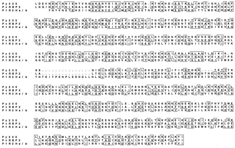
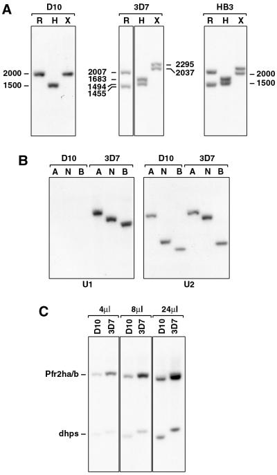
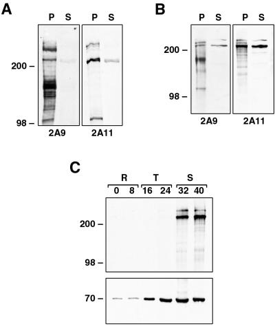
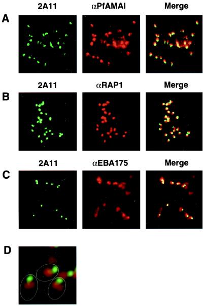
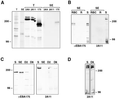
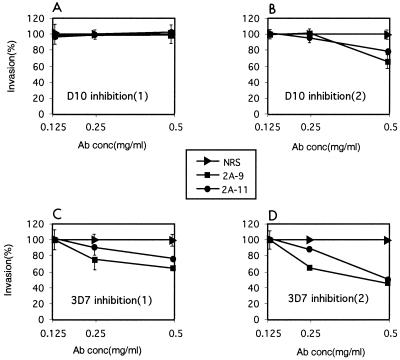
Plasmodium falciparum infections can be fatal, while P. vivax infections usually are not. A possible factor involved in the greater virulence of P. falciparum is that this parasite grows in red blood cells (RBCs) of all maturities whereas P. vivax is restricted to growth in reticulocytes, which represent only approximately 1% of total RBCs in the periphery. Two proteins, expressed at the apical end of the invasive merozoite stage from P. vivax, have been implicated in the targeting of reticulocytes for invasion by this parasite. A search of the P. falciparum genome databases has identified genes that are homologous to the P. vivax rbp-1 and -2 genes. Two of these genes are virtually identical over a large region of the 5' end but are highly divergent at the 3' end. They encode high-molecular-mass proteins of >300 kDa that are expressed in late schizonts and localized to the apical end of the merozoite. To test a potential role in merozoite invasion of RBCs, we analyzed the ability of these proteins to bind to mature RBCs and reticulocytes. No binding to mature RBCs or cell preparations enriched for reticulocytes was detected. We identified a parasite clone that lacks the gene for one of these proteins, showing that the gene is not required for normal in vitro growth. Antibodies to these proteins can inhibit merozoite invasion of RBCs.


Figures

FIG. 1
FIG. 1
Comparison of the 500-amino-acid region conserved between PvRBP-2 and Py235 and its sequence in the P. falciparum homologues PfR2Ha and -Hb. Accession numbers for the sequence data shown: Py235, L27838; Pvrbp-2, Q00799; Pfr2ha/hb (AL049181, AF312916, and AF312917). Sequences were aligned using CLUSTAL V software. Boxes represent positions which have >50% identity; dots represent spaces inserted into the sequence to provide optimal homology.
FIG. 2
FIG. 2
3D7 parasites have both Pfr2ha and Pfr2hb genes, while D10 has only Pfr2ha. (A) Schematic representation of the Pfr2hb gene in 3D7 parasites. The short signal peptide (exon 1) is followed by an intron and then exon 2. The C1 probe common to both Pfr2ha and Pfr2hb was amplified from D10 gDNA using primers 5′-ACAGGAAATATGTGAAAAACGG-3′ and 5′-TTATTATTATTAGTGTTTTTAC-3′. The C2 probe common to both Pfr2ha and Pfr2hb was amplified from 3D7 gDNA using primers 5′-CACCAAGATCCTTTATATCA-3′ and 5′-CTTAATATAAATAATATTATGAAT-3′. The U1 probe, unique to the Pfr2hb gene, was amplified from 3D7 gDNA using primers 5′-GAATTGATAGTACTGACCAACGT-3′ and 5′-CTTCATTTTCATCAAACACAATTTC-3′. A region of 2,312 bp bounded by RsaI (R) and XmnI (X) sites is shown expanded below the Pfr2hb gene. The HinfI (H) site is shown together with fragment sizes in base pairs. (B) Schematic representation of the Pfr2ha gene in D10 and 3D7 parasites. The C1 and C2 probes are as in panel A. The U2 probe was amplified from 3D7 gDNA using primers 5′-TAAACTAGAATCTGATATGGTGA-3′ and 5′-GTCATCTTTTTTTTCTTTAGATGT-3′. A region of 2,037 bp bounded by RsaI and XmnI sites is shown expanded below the Pfr2ha gene. (C) Schematic representation of the PfR2Ha and -Hb proteins in 3D7 and the PfR2Ha protein in D10 parasites. The three sequences are presumed to be nearly identical (see Results) but differ markedly from amino acid (aa) 2776 onward even though they are structurally similar, with a putative transmembrane domain (TM) and a short cytoplasmic tail at the C terminus. The unique regions are shown as diagonally hatched in R2Hb and horizontally hatched in R2Ha. The 500-amino-acid region showing some conservation in PvRBP-2 and Py235 as shown in Fig. 1 is indicated. The DNA corresponding to approximately 1,100 amino acids at the C terminus of the 3D7 r2ha and r2hb genes and approximately 800 amino acids at the C terminus of the D10 r2hb gene was sequenced. This encompassed the regions to which the 2A9 and 2A11 antibodies were made. For PCR amplification of the D10 and 3D7 r2ha genes, primers P1 (5′-AATTACGTGAATTGTCTACGGC-3′) and P2 (5′-GTCATCTTTTTTTTCTTTAGATGTTATC-3′) were used. For amplification of the 3D7 r2hb gene, primers P1 and P3 (5′-AAACAACATGATCATACGCATTG-3′) were used. The approximate locations of primers P1, P2, and P3 are shown. The PCR products were fully sequenced using internal primers. The amino acid differences within the regions of the three genes which are nearly identical are indicated by asterisks. The 5′-most change (amino acid 2546) is A (Ala) in 3D7 R2Ha but D (Asp) in the other proteins. The next change (position 2635) is E (Glu) in both R2Ha proteins but K (Lys) in 3D7 R2Hb. The 3′-most change (beginning at position 2719) is EEELRKK in 3D7 R2Ha but EALKKQ in the other proteins. The portions of PfR2Ha and -Hb used for production of rabbit antibodies 2A9 and 2A11 are also shown. The lengths of the signal sequence (S) and transmembrane domain (TM) are not shown to scale. The checkered shading represents the 500-amino-acid conserved region. The diagonal and horizontal shaded regions represent the unique regions of PfR2Hb and PfR2Ha, respectively.
FIG. 3
FIG. 3
D10 parasites lack the Pfr2hb gene. (A) D10, 3D7, and HB3 gDNAs were digested with RsaI (R), HinfI (H), or XmnI (X), blotted to HybondN, and probed with the C2 probe. Sizes are in base pairs. (B) D10 and 3D7 gDNAs were digested with AccI (A), NsiI (N), or BstYI (B), blotted to HybondN, and probed with the Pfr2hb-specific probe U1 or the Pfr2ha-specific probe U2. (C) D10 and 3D7 gDNAs were double digested with NsiI/BglII, blotted to HybondN, and probed with a mixture of the single-copy gene dhps (21) and the C1 probe. Either 4, 8, or 24 μl of the digest was electrophoresed on the agarose gel. The dhps fragment was amplified from D10 gDNA using primers 5′-AAGATTAAATTTTCTTG-3′ and 5′-ATATAGAATTGTTACTTTTGTATA-3′. The copy number of the Pfr2ha and -hb genes in 3D7 relative to D10 was determined using a PhosphorImager (Molecular Dynamics).
FIG. 4
FIG. 4
(A and B) Specificities of anti-PfRH antibodies. Two different anti-PfRH antibodies show similar specificities on HB3 (A) and D10 (B) parasites. For production of parasite pellets (P), cultures were sorbitol synchronized, cultured until the late-schizont stage, and saponin lysed, and then the pellet was resuspended in reducing sample buffer. Production of the EBA175 supernatant (S) is as described in Materials and Methods. Protein samples were separated by SDS-PAGE, transferred to nitrocellulose, and probed with both the 2A9 and 2A11 antibodies. (C) Protein samples from synchronized D10 parasites taken at 8-h intervals were separated by SDS-PAGE, transferred to nitrocellulose, and probed with both the 2A11 (top) and anti-Pfhsp70 (bottom) antibodies. During development, parasites transform from rings (R), through trophozoites (T), and to schizonts (S). Sizes are in kilodaltons.
FIG. 5
FIG. 5
PfR2Ha and -Hb do not colocalize with either PfAMA1, EBA175, or PfRAP1 by IFA analysis. Smears of free merozoites following schizont rupture of the D10 parasite are shown. Parasites were reacted with mouse antibodies to PfAMA1 (A), PfRAP1 (B), EBA175 (C), or PfRAP1 together with a rabbit antibody to PfR2Ha/Hb (2A11) (D). For images in the first column, the second antibody used was a sheep FITC-labeled anti-rabbit antibody; for images in the second column, the second antibody used was a goat rhodamine-labeled anti-mouse antibody; the Merge column shows the red and green images overlaid. Magnifications: A to C, ×1,000; D, ×3,000. The outline of three merozoites is shown in panel D.
FIG. 6
FIG. 6
Do PfR2Ha and -Hb bind human RBCs? (A) 35S-labeled supernatant from D10 parasites was either immunoprecipitated directly with the anti-PfRh antibodies 2A9 and 2A11 or anti-EBA175 antibody or immunoprecipitated with the same antibodies after proteins were bound to RBCs and eluted with 300 mM NaCl. Total labeled proteins (T) or proteins eluted from RBCs (SE [salt eluted]) together with immunoprecipitated proteins were separated by SDS-PAGE and then detected by standard fluorographic methods. (B) Unlabeled supernatant from 3D7 parasites (200 μl) was bound to equal numbers of Percoll-purified reticulocytes enriched to 10% (R) and RBCs depleted of reticulocytes (RBC) by passage over Percoll. Bound proteins were eluted with 300 mM NaCl, separated by SDS-PAGE, transferred to nitrocellulose, and then probed with antibodies to EBA175 and PfRh (2A11). Two microliters of the supernatant (S) was also run as a positive control. SE, salt eluted. (C) Unlabeled supernatant from 3D7 parasites (100 μl) was bound to 20 μl of RBC and then eluted with 300 mM NaCl (SE). The supernatant was further depleted either two times (D2) or eight times (D8) by repeated addition of 20 μl of RBC for 30 min. Either 2 μl of the original supernatant (S) or between 2.5 and 3.2 μl (to allow for volume increases during repeated RBC additions) of the depleted supernatants, together with the salt-eluted proteins, was separated by SDS-PAGE, transferred to nitrocellulose, and then probed with antibodies to EBA175 and PfRh (2A11). (D) Unlabeled supernatant from 3D7 parasites (100 μl) which had been ultracentrifuged at 100,000 for 30 min was bound to 20 μl of RBC. The supernatant was depleted six times (D6) by repeated addition of 20 μl of RBC for 30 min. Either 2 μl of the original supernatant (S) or 2.6 μl of the depleted supernatant was separated by SDS-PAGE, transferred to nitrocellulose, and probed with the 2A11 antibody.
FIG. 7
FIG. 7
Antibodies to PfR2Ha and -Hb result in some invasion inhibition. Purified schizonts from D10 and 3D7 parasites were plated in human erythrocytes to determine the ability of released merozoites to invade in the presence or absence of protein G-purified 2A9 and 2A11 antibodies (0.125, 0.25, and 0.50 mg/ml) for each parasite, results of two independent experiments done in duplicate are shown. Invasion in the presence of 0.125, 0.25 and 0.50 mg of protein G-purified normal NRS per ml was adjusted to 100%. Percent invasion was determined microscopically by counting at least 1,000 RBCs. The parasitemias at the end of the experiment were between 9 and 12% for the control wells containing NRS. Standard errors are shown except those less than 4% (e.g., panel D); the standard errors ranged from 0 to 14%.

References

    1. Bianco A E, Favaloro J M, Burkot T R, Culvenor J G, Crewther P E, Brown G V, Anders R F, Coppel R L, Kemp D J. A repetitive antigen of Plasmodium falciparum that is homologous to heat shock protein 70 of Drosophila melanogaster. Proc Natl Acad Sci USA. 1986;83:8713–8717. - PMC - PubMed
    1. Borre M B, Owen C A, Keen J K, Sinha K A, Holder A A. Multiple genes code for high-molecular-mass rhoptry proteins of Plasmodium yoelii. Mol Biochem Parasitol. 1995;70:149–155. - PubMed
    1. Dolan S A, Proctor J L, Alling D W, Okubo Y, Wellems T E, Miller L H. Glycophorin B as an EBA-175 independent Plasmodium falciparum receptor of human erythrocytes. Mol Biochem Parasitol. 1994;64:55–63. - PubMed
    1. Freeman R R, Trejdosiewicz A J, Cross G A. Protective monoclonal antibodies recognising stage-specific merozoite antigens of a rodent malaria parasite. Nature. 1980;284:366–368. - PubMed
    1. Galinski M R, Medina C C, Ingravallo P, Barnwell J W. A reticulocyte-binding protein complex of Plasmodium vivax merozoites. Cell. 1992;69:1213–1226. - PubMed

Publication types

Associated data

LinkOut - more resources