. 2001 Jan;107(2):225-34.
doi: 10.1172/JCI10832.

LPS-binding protein circulates in association with apoB-containing lipoproteins and enhances endotoxin-LDL/VLDL interaction

Affiliations

LPS-binding protein circulates in association with apoB-containing lipoproteins and enhances endotoxin-LDL/VLDL interaction

A C Vreugdenhil et al. J Clin Invest. 2001 Jan.

Abstract

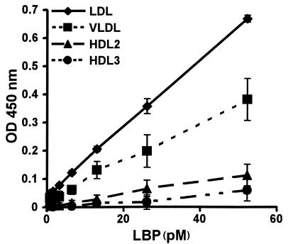
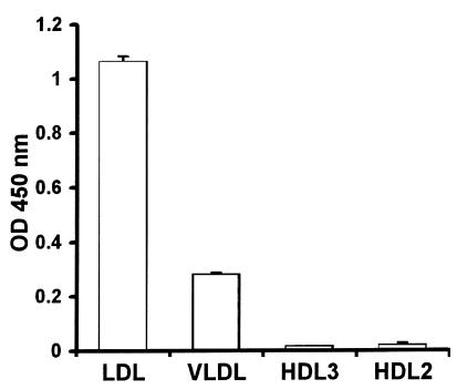
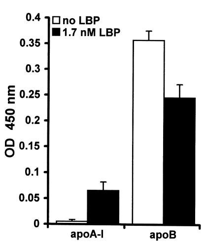
LPS-binding protein (LBP) and serum lipoproteins cooperate in reducing the toxic properties of LPS. In the present study, we demonstrate that LBP circulates in association with LDL and VLDL in healthy persons. ApoB was found to account at least in part for the interaction of LBP with LDL and VLDL. Although LBP interacted with purified apoA-I in vitro, no association of LBP with apoA-I or HDL was found in serum. Consistent with the observed association of LBP with LDL and VLDL, these lipoproteins also were demonstrated to be the predominant LPS-binding lipoproteins. Most interestingly, the association of LBP with LDL and VLDL strongly enhanced the capacity of these lipoproteins to bind LPS. Because this function of LBP is of utmost importance during infection, the association of LBP and LPS with lipoproteins was also studied in serum from septic patients. In septic serum containing high LBP levels and a markedly altered lipoprotein spectrum, most of the LBP is associated with LDL and VLDL, although some LBP appeared to circulate free from lipoproteins. Also in this serum, LPS was found to bind predominantly to LDL and VLDL. The observed binding of LBP and LPS to LDL and VLDL, as well as the LBP-dependent incorporation of LPS into these lipoproteins, emphasizes a crucial role for circulating LBP-LDL/VLDL complexes in the scavenging of LPS.


Figures

Figure 1
Figure 1
Distribution profile of LBP among lipoproteins in serum of healthy persons. Serum from three healthy persons (lanes 1–3) and LBP-depleted serum (lane 4) were subjected to agarose gel electrophoresis, blotted, and probed with specific antibodies to human LBP, apoA-I, and apoB. LBP colocalizes with apoB in the β mobility region and not with apoA-I.
Figure 2
Figure 2
Effect of LDL and VLDL on the electrophoretic mobility of purified LBP. Purified LBP was preincubated with PBS and isolated LBP-free HDL, VLDL, or LDL (0.1, 0.035, and 0.5 mg cholesterol/ml respectively). Agarose gel electrophoresis of purified LBP and the preincubated fractions was followed by Western blot analysis of LBP using a specific antibody. Purified LBP migrates in between the β and α mobility regions. Preincubation of LBP with isolated VLDL or LDL shifts the migration of LBP to the β mobility region.
Figure 3
Figure 3
Presence of apoB in LBP-containing lipoproteins. LBP-containing lipoproteins were captured from serum by applying serum to 96-well plates coated with an mAb against LBP (diamonds) followed by extensive washing of the plates. As a control serum was also applied to noncoated plates (squares) or plates coated with an aspecific antibody (rat anti-murine TNF-R75) (triangles). Presence of apoB in the captured lipoproteins was detected by addition of a peroxidase-labeled mAb against apoB and is expressed as mean ± SD of the OD 450 nm of four wells.
Figure 4
Figure 4
Association of LBP with different lipoprotein classes. Lipoproteins isolated from human serum by ultracentrifugation, free of LBP, and standardized for cholesterol concentration were immobilized to 96-well plates and incubated with biotinylated LBP. Bound LBP was detected by peroxidase-conjugated streptavidin and TMB. Binding of LBP to the lipoproteins is expressed as mean ± SD of the OD 450 nm of four wells after correction for background. LDL and VLDL display high LBP-binding capacity in contrast to HDL.
Figure 5
Figure 5
Distribution profile of LPS among lipoproteins in serum of healthy persons. Serum of three healthy donors and LBP-depleted serum were preincubated with biotinylated LPS. Agarose gel electrophoresis of the sera was followed by Western blotting. Biotinylated LPS was detected using peroxidase-conjugated streptavidin and a chemiluminescent substrate. LPS incubated with serum is recovered in the β mobility region.
Figure 6
Figure 6
Association of LPS with different lipoprotein classes. Lipoproteins isolated from human serum by ultracentrifugation, free of LBP, and standardized for cholesterol concentration were immobilized to 96-well plates followed by incubation with biotinylated LPS. Binding of LPS to the lipoproteins was detected by peroxidase-conjugated streptavidin and TMB and expressed as mean ± SD of the OD 450 nm of four wells after correction for background. LDL and to a lesser extent VLDL display high LPS-binding capacity in contrast to HDL.
Figure 7
Figure 7
LBP associated with LDL and VLDL enhances the interaction of LPS with lipoproteins. Plates were coated with isolated LDL (2 mg cholesterol/ml) or VLDL (14 mg cholesterol/ml). The immobilized lipoproteins were preincubated with a concentration range of LBP overnight at 37°C. Unbound LBP was removed by washing the plates, and biotinylated LPS was added to the LBP-lipoprotein complexes. Plates were washed to remove unbound LPS and bound LPS was detected using peroxidase-conjugated streptavidin and TMB. Binding of LPS to the lipoproteins is expressed as mean ± SD of the OD 450 nm of four wells after correction for background. LBP associated with LDL and VLDL enhances the binding of LPS dose-dependently.
Figure 8
Figure 8
Association of LBP and LPS with apoB and apoA-I. (a) Binding of biotin-labeled LPS to apolipoproteins was evaluated. To this end, plates were coated with a concentration range of apoB and apoA-I, and biotin-labeled LPS was added. Bound LPS was detected by peroxidase-conjugated streptavidin and TMB and expressed as mean ± SD of the OD 450 nm of four wells after correction for background. (b) Binding of biotin-labeled LBP to immobilized apoB (25 nM) or apoA-I (100 nM) was detected by peroxidase-conjugated streptavidin and TMB and expressed as mean ± SD of the OD 450 nm of four wells after correction for background. (c) The relative affinities of LBP for apoB and apoA-I were evaluated. Biotinylated LBP was added to plates coated with apoB (25 nM). Inhibition of this interaction of LBP with apoB by apoA-I and apoB was studied by adding concentration ranges of apoA-I and apoB together with LBP. Bound LBP was detected by peroxidase-conjugated streptavidin and TMB and expressed as mean ± SD of the OD 450 nm of three wells after correction for background.
Figure 9
Figure 9
LBP enhances binding of LPS to apoA-I and reduces binding of LPS to apoB. Plates were coated with apoA-I (25 nM) or apoB (100 nM). A total of 1.7 nM of LBP was added together with biotinylated LPS, followed by peroxidase-conjugated streptavidin and TMB. Binding of LBP to the apolipoproteins is expressed as mean ± SD of the OD 450 nm of four wells after correction for background. The binding of LPS to apoA-I is markedly enhanced by LBP, whereas LBP reduces the binding of LPS to apoB.
Figure 10
Figure 10
Distribution profile of LBP and LPS in serum of septic patients. Agarose gel electrophorese of serum from four septic patients (lanes 1–4) was followed by Western blot analyses using specific antibodies for LBP, and apoB. Agarose gel electrophorese of serum from three septic patients (lanes 1–3) and a serum pool of healthy persons (lane 4) was followed by Western blot analyses using specific antibodies for apoA-I. LBP predominantly colocalizes with apoB. An additional band compared with normal serum is observed in all subjects between the β and α regions, and is most explicit in subject 3. Preincubation of LPS with the sera followed by Western blot analyses demonstrates the distribution of LPS among lipoproteins during an acute-phase response. LPS colocalizes with LBP and apoB in the β region under these conditions.

References

    1. Read TE, et al. The protective effect of serum lipoproteins against bacterial lipopolysaccharide. Eur Heart J. 1993;14:125–129. - PubMed
    1. Feingold KR, et al. Role for circulating lipoproteins in protection from endotoxin toxicity. Infect Immun. 1995;63:2041–2046. - PMC - PubMed
    1. Flegel WA, Wölpl A, Männel DN, Northoff H. Inhibition of endotoxin-induced activation of human monocytes by human lipoproteins. Infect Immun. 1989;57:2237–2245. - PMC - PubMed
    1. Ulevitch RJ, Johnston AR, Weinstein DB. New function for high density lipoproteins. J Clin Invest. 1979;64:1516–1524. - PMC - PubMed
    1. Van Lenten BJ, Fogelman AM, Haberland ME, Edwards PA. The role of lipoproteins and receptor-mediated endocytosis in the transport of bacterial lipopolysaccharide. Proc Natl Acad Sci USA. 1986;83:2704–2708. - PMC - PubMed

Publication types