. 2001 Jan;107(2):R1-7.
doi: 10.1172/JCI11642.

Prevention of cardiomyopathy in mouse models lacking the smooth muscle sarcoglycan-sarcospan complex

Affiliations

Prevention of cardiomyopathy in mouse models lacking the smooth muscle sarcoglycan-sarcospan complex

R D Cohn et al. J Clin Invest. 2001 Jan.

Abstract

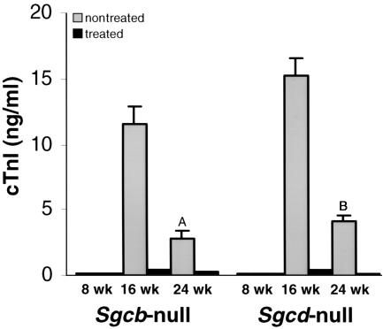
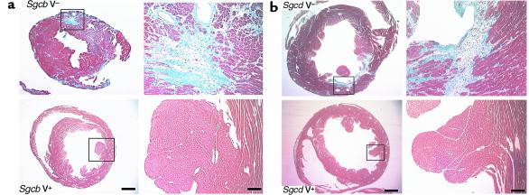
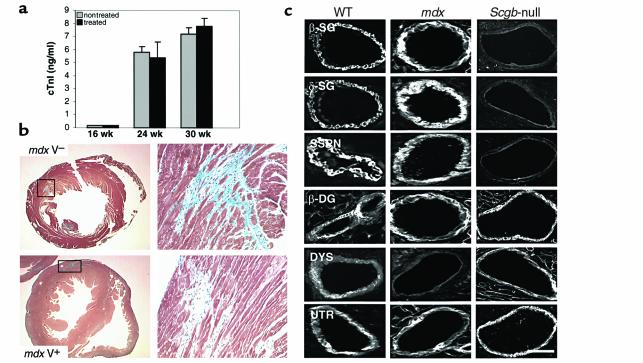
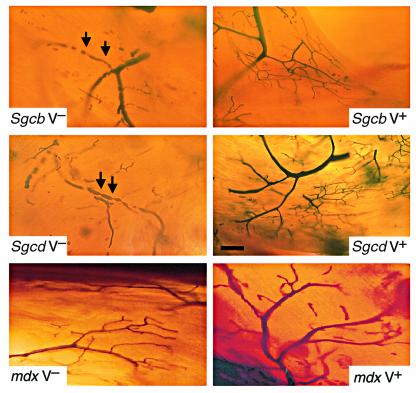
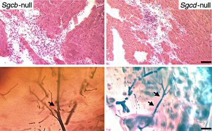
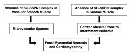
Cardiomyopathy is a multifactorial disease, and the dystrophin-glycoprotein complex has been implicated in the pathogenesis of both hereditary and acquired forms of the disease. Using mouse models of cardiomyopathy made by ablating genes for components of the sarcoglycan complex, we show that long-term treatment with verapamil, a calcium channel blocker with vasodilator properties, can alleviate the severe cardiomyopathic phenotype, restoring normal serum levels for cardiac troponin I and normal cardiac muscle morphology. Interruption of verapamil treatment leads again to vascular dysfunction and acute myocardial necrosis, indicating that predilection for cardiomyopathy is a continuing process. In contrast, verapamil did not prevent cardiac muscle pathology in dystrophin-deficient mdx mice, which neither show a disruption of the sarcoglycan complex in vascular smooth muscle nor vascular dysfunction. Hence, our data strongly suggest that pharmacological intervention with verapamil merits investigation as a potential therapeutic option not only for patients with sarcoglycan mutations, but also for patients with idiopathic cardiomyopathy associated with myocardial ischemia not related to atherosclerotic coronary artery disease.


Figures

Figure 1
Figure 1
Levels of cTnI in verapamil-treated versus untreated Sgcb- and Sgcd-null mice. Note the significant elevation of cTnI in the untreated group of Sgcb- and Sgcd-null mice. Data are represented as mean plus or minus SD. ASgcb-null treated versus untreated P < 0.002; BSgcd-null treated versus untreated P < 0.001.
Figure 2
Figure 2
Morphological analysis of cardiac muscle of Sgcb- and Sgcd-null mice. The untreated mice (V–) show extensive areas of fibrosis in the heart (left panels). In contrast, most of the cardiac muscle of verapamil-treated mice (V+) is free of any morphological alterations (right panels). Bar, 1.5 mm and 75 μm, respectively. (a) Sgcb-null mice. (b) Sgcd-null mice.
Figure 3
Figure 3
Verapamil does not prevent cardiomyopathy in dystrophin-deficient mdx mice. (a) Levels of cTnI in verapamil-treated versus untreated mdx mice. Both groups of mice show elevations of cTnI levels in the serum. (b) Histological analysis of hearts from mdx mice after treatment with verapamil reveals areas of fibrosis in both groups of mice. (c) Immunohistochemical expression of β-sarcoglycan, δ-sarcoglycan, sarcospan, and β-dystroglycan is preserved in vascular smooth muscle of coronary arteries of mdx mice. In contrast, the SG-SSPN complex is absent in vascular smooth muscle of Sgcb-null mice. Note that utrophin is expressed at similar levels in wild-type, mdx, and Sgcb-null mice. β-SG, β-sarcoglycan; δ-SG, δ-sarcoglycan; β-DG, β-dystroglycan; DYS, dystrophin; and UTR, utrophin. Bar, 20 μm.
Figure 4
Figure 4
Perfusion of coronary arteries of verapamil-treated and untreated mice. Transillumination of Microfil-perfused coronary arteries in untreated mice shows multiple constrictions (arrows) with pre- and poststenotic dilations, as well as narrow vessels with a serrated, rather than a smooth, contour (upper and middle left panels). In contrast, coronary vessels of treated Sgcb- and Sgcd-null mice (upper and middle right panels) reveal smoothly tapered vessels without signs of constriction and/or focal narrowing. Note the normal vascular perfusion of treated and untreated mdx mice (lower panel). Bar, 40 μm.
Figure 5
Figure 5
Interruption of verapamil treatment leads to vascular dysfunction and myocardial necrosis. Analysis of cardiac muscle morphology and vascular perfusion 10 days after interruption of 16 weeks of verapamil therapy exhibits the development of focal necrotic lesions and microvascular constrictions in Sgcb- and Sgcd-null mice. Bar, 75 μm and 40 μm, respectively.
Figure 6
Figure 6
Pathogenesis of cardiomyopathy in LGMD 2E/2F. The flow chart represents the current understanding of the pathogenesis of cardiomyopathy due to primary mutations within the β- or δ-sarcoglycan gene. Absence of SG-SSPN complex in vascular smooth muscle leads to vascular dysfunction in the form of microvascular spasms. In addition, loss of the SG-SSPN complex in cardiac muscle renders the heart susceptible to intermittent ischemic-like events, which eventually leads to the development of focal myocardial necrosis and severe cardiomyopathy.

Comment in

References

    1. Schocken DD, Arrieta MI, Leaverton PE, Ross EA. Prevalence and mortality rate of congestive heart failure in the United States. J Am Coll Cardiol. 1992;20:301–306. - PubMed
    1. Chen J, Chien KR. Complexity in simplicity: monogenic disorders and complex cardiomyopathies. J Clin Invest. 1999;103:1483–1485. - PMC - PubMed
    1. Nigro G, Comi LI, Politano L, Bain RJ. The incidence and evolution of cardiomyopathy in Duchenne muscular dystrophy. Int J Cardiol. 1990;26:271–277. - PubMed
    1. Badorff C, et al. Enteroviral protease 2A cleaves dystrophin: evidence of cytoskeletal disruption in an acquired cardiomyopathy. Nat Med. 1999;5:320–326. - PubMed
    1. Coral-Vazquez R, et al. Disruption of the sarcoglycan-sarcospan complex in vascular smooth muscle: a novel mechanism for cardiomyopathy and muscular dystrophy. Cell. 1999;98:465–474. - PubMed

Publication types

MeSH terms