. 2001 Jan;107(2):R9-R14.
doi: 10.1172/JCI10559.

Delayed wound repair and impaired angiogenesis in mice lacking syndecan-4

Affiliations

Delayed wound repair and impaired angiogenesis in mice lacking syndecan-4

F Echtermeyer et al. J Clin Invest. 2001 Jan.

Abstract

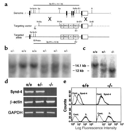
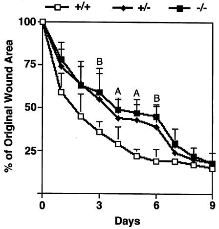
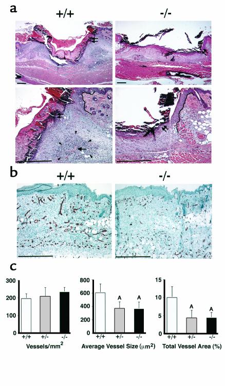
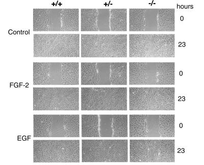
The syndecans make up a family of transmembrane heparan sulfate proteoglycans that act as coreceptors with integrins and growth factor tyrosine kinase receptors. Syndecan-4 is upregulated in skin dermis after wounding, and, in cultured fibroblasts adherent to the ECM protein fibronectin, this proteoglycan signals cooperatively with beta1 integrins. In this study, we generated mice in which the syndecan-4 gene was disrupted by homologous recombination in embryonic stem cells to test the hypothesis that syndecan-4 contributes to wound repair. Mice heterozygous or homozygous for the disrupted syndecan-4 gene are viable, fertile, and macroscopically indistinguishable from wild-type littermates. Compared with wild-type littermates, mice heterozygous or homozygous for the disrupted gene have statistically significant delayed healing of skin wounds and impaired angiogenesis in the granulation tissue. These results indicate that syndecan-4 is an important cell-surface receptor in wound healing and angiogenesis and that syndecan-4 is haplo-insufficient in these processes.


Figures

Figure 1
Figure 1
Generation and characterization of syndecan-4 recombinant mice. (a) Structure of the murine syndecan-4 gene, the targeting vector, and the targeted allele. Exons are numbered 1 through 5. This organization is similar to that reported by others (25). Sa, SacI; Sp, SpeI; Av, AvrII; EV, EcoRV; B, BamHI; Xh, XhoI; Sn, SnaBI. SA, splice acceptor; IRES, internal ribosomal entry site; LACZ, β-galactosidase; NEO, neomycin resistance. (b) Southern blot analysis of ES clones. (c) Southern blot analysis of mice derived from matings between syndecan-4+/– mice. Wild-type (14.1 kb) and targeted alleles (12 kb) are detected with the 5′ probe. (d) RT-PCR of mRNA results in a syndecan-4–specific 524-bp band from syndecan-4+/+ and syndecan-4+/– cells but not from syndecan-4–/– cells. mRNA for both β-actin and GAPDH is present in extracts of cells of all three genotypes. (e) Flow cytometric analysis of cell-surface syndecan-4 on skin fibroblasts. Syndecan-4 (Synd-4) is detected on syndecan-4+/+ cells (upper left) but not on syndecan-4–/– cells (upper right). β1 Integrins (β1) are expressed on cells of both genotypes (lower panels). Secondary antibodies alone were used as control (Cont).
Figure 2
Figure 2
Delayed wound healing in mice homozygous (–/–) or heterozygous (+/–) for the recombinant syndecan-4 allele compared with wild-type (+/+) littermates. A statistically significant delay is seen between days 3 and 6 after wounding. Data are expressed as means ± SEM (n = 20). AP < 0.05. BP < 0.01.
Figure 3
Figure 3
(a) Histological analysis of wound healing in syndecan-4+/+ (left panels) and syndecan-4–/– (right panels) mice at 3 days after wounding. The higher magnification in the bottom panels is of the region in the upper right edge of the wounds in the respective top panels. Double arrows: margin of epithelium; single arrow: blood vessel in dense granulation tissue (arrowheads); solid lines in bottom panels: wound edge. Bar, 200 μm. (b) Immunohistochemical examination of wounds 6 days after wounding for the endothelial cell marker CD31 in syndecan-4+/+ (left) and in syndecan-4–/– (right) mice. Bar, 100 μm. (c) Quantitative analysis of angiogenesis. The numbers of vessels per unit area are the same for the three genotypes (left). Average vessel size (middle) and total vessel area (right) in the wounds of syndecan-4–/– or syndecan-4+/– mice are significantly different from those of syndecan-4+/+ mice. Data are expressed as means ± SEM (n = 8). AP < 0.05.
Figure 4
Figure 4
In vitro migration analysis of syndecan-4 recombinant fibroblasts. Wild-type and heterozygous fibroblasts fill in a space created in an in vitro wound healing model completely by 23 hours in contrast to syndecan-4–/– cells. Fibroblasts were grown to confluency in 10% FBS and then placed in 2% FBS overnight. The in vitro wound assays were performed on cultures of all three genotypes in control medium (DMEM containing 2% FBS) or medium supplemented with 25 ng/ml of FGF-2 or 25 ng/ml of EGF. The wound closure was monitored over time and terminated upon complete closure of the wounds by the syndecan-4+/+ fibroblasts.

References

    1. Clark RA. Biology of dermal wound repair. Dermatol Clin. 1993;11:647–666. - PubMed
    1. Kyriakides TR, Tam JWY, Bornstein P. Accelerated wound healing in mice with a disruption of the thrombospondin 2 gene. J Invest Dermatol. 1999;113:782–787. - PubMed
    1. Streit M, et al. Thrombospondin-1 suppresses wound healing and granulation tissue formation in the skin of transgenic mice. EMBO J. 2000;19:3272–3282. - PMC - PubMed
    1. Ortega S, Ittmann M, Tsang SH, Ehrlich M, Basilico C. Neuronal defects and delayed wound healing in mice lacking fibroblast growth factor 2. Proc Natl Acad Sci USA. 1998;95:5672–5677. - PMC - PubMed
    1. Burridge K, Chrzanowska-Wodnicka M. Focal adhesions, contractility, and signaling. Annu Rev Cell Dev Biol. 1996;12:463–518. - PubMed

Publication types