Comparative Study
. 2001 Jan;107(2):R15-22.
doi: 10.1172/JCI11540.

IFN-gamma action in the media of the great elastic arteries, a novel immunoprivileged site

Affiliations
Comparative Study

IFN-gamma action in the media of the great elastic arteries, a novel immunoprivileged site

A J Dal Canto et al. J Clin Invest. 2001 Jan.

Abstract

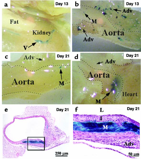
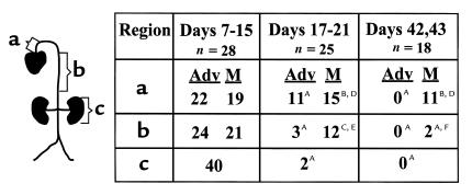
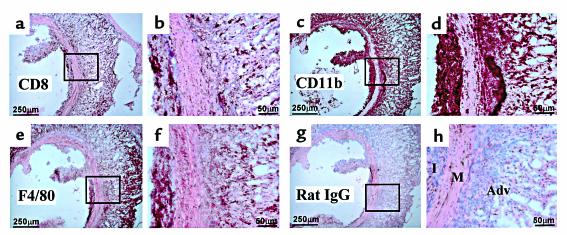
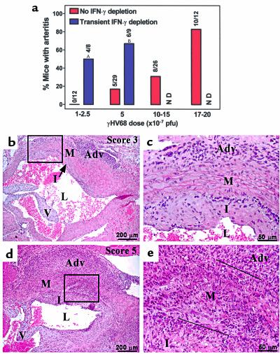
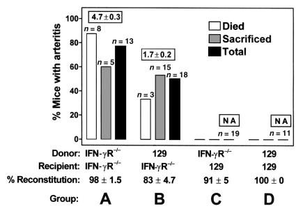
Infection of medial smooth muscle cells with gamma-herpesvirus 68 (gammaHV68) causes severe chronic vasculitis that is restricted to the great elastic arteries. We show here that persistence of disease in the great elastic arteries is (a) due to inefficient clearance of viral infection from this site compared with other organs or other vascular sites, and (b) associated with failure of T cells and macrophages to enter the virus-infected elastic media. These findings demonstrate immunoprivilege of the media of the great elastic arteries. We found that IFN-gamma acted on somatic cells during acute infection to prevent the establishment of medial infection and on hematopoietic cells to determine the severity of disease in this site. The immunoprivileged elastic media may provide a site for persistence of pathogens or self antigens leading to chronic vascular disease, a process regulated by IFN-gamma actions on both somatic and hematopoietic cells. These concepts have significant implications for understanding immune responses contributing to or controlling chronic inflammatory diseases of the great vessels.


Figures

Figure 1
Figure 1
Localization of γHV68 vascular infection over time. IFNγR–/– mice were infected with either M1.LacZ or γHV68 v-cyclin.LacZ. The aorta and its major branches were resected and stained en bloc for β-gal activity. Infection with wild type γHV68 resulted in no comparable staining.
Figure 2
Figure 2
Quantitation of clearance of γHV68 from different vascular structures over time. IFN-γR–/– mice were infected and analyzed as in Figure 1 at days 7, 9, 11, 13, 15, 17, 19, 21, 42, and 43 after infection. n, number of mice. The number of aortas with blue staining in the adventitia (Adv) or media (M) is reported for the aortic base (a) or the descending aorta (b), which was representative of the entire aorta. Staining of a peripheral vessel on the posterior surface of both kidneys is also reported (c). For staining at late times compared with staining at days 7–15, AP < 0.0001; BP > 0.05; CP = 0.04. For medial clearance compared with adventitial clearance, DP < 0.0001; EP = 0.006; FP = 0.1.
Figure 3
Figure 3
Localization of inflammatory cells in arteritic lesions. Shown are low- and high-power views of immunohistochemistry on parallel aortic frozen sections from an IFN-γR–/– mouse infected with γHV68 4 weeks after infection. Dark-brown staining represents Ab binding. (a and b) CD8 T cells are present in the intima and adventitia, but not the media. Similar staining was observed with an Ab to CD4 (not shown). (c and d) Staining for CD11b shows Mac1-positive cells (macrophages and neutrophils) in all three layers of the aorta. (e and f) Staining for F4/80-positive macrophages is limited to the intima and adventitia. (g and h) Negative control with rat IgG. The area of highest background staining is shown in h for comparison with specific staining in panels b, d, and f. I, intima; M, media; Adv, adventitia.
Figure 4
Figure 4
Incidence and severity of chronic elastic arteritis in wild-type mice infected with high doses of γHV68 with or without transient depletion of IFN-γ. Mice were infected with the indicated doses of γHV68 with or without depletion of IFN-γ and evaluated for arteritis on H&E-stained sections 5.5 to 10 weeks after infection. (a) The incidence of arteritis in various groups is presented. Seven of 19 mice infected with 17 to 20 × 107 PFU γHV68 died within 7 days. Twelve of 14 mice infected with 34 to 50 × 107 PFU died within 7 days. Data for transient depletions are from two independent experiments. Data for no depletions are from 4–5 independent experiments. AP = 0.0062; BP = 0.0043. (b) Lesion at the aortic base of a 129Ev/Sv mouse sacrificed 10 weeks after infection with 108 PFU γHV68. Lesions in transiently IFN-γ–depleted 129Ev/Sv mice have similar histology (lesion scores for 25 mice from both groups combined = 2.0 ± 0.2; P < 0.0001 for scores compared with IFN-γR–/– mice, P = 0.001 compared with chronically depleted wild-type mice). (c) High-power view of boxed region in b. (d) Lesion at the aortic base of a chronically IFN-γ–depleted 129Ev/Sv mouse sacrificed 6 weeks after infection with 5 × 10⁷ PFU γHV68 (lesion scores for four mice = 4.0 ± 0.7). Arteritis in IFN-γR–/– mice has similar histology (lesion scores for 16 mice = 5.0 ± 0). (e) High-power view of boxed region in d. The black lines show the boundaries of the media. Adv, adventitia; M, media; I, intima; L, lumen; V, aortic valve. ND, not determined.
Figure 5
Figure 5
IFN-γ prevents chronic elastic arteritis by effects on somatic cells but regulates the nature of the pathology by effects on hematopoietic cells. Reciprocal bone marrow reconstitutions were performed between IFN-γR–/– and 129Ev/Sv mice, and arteritis was evaluated for 12 weeks after γHV68 infection. Numbers above bars represent the number of mice within a group. Boxed numbers represent average lesion scores for a group. Data are pooled from four independent experiments. For incidence of disease, P = 0.0001 comparing group A with D, and P = 0.0004 comparing group B with C. For severity of disease, P < 0.0001 comparing groups A and B. NA, lesion scores not applicable.
Figure 6
Figure 6
IFN-γ inhibits γHV68 infection of primary aortic cells. IFN-γ–treated and untreated intimal/adventitial and medial cultures were evaluated 3 days after infection. In c and d nuclei were stained blue, muscle actin was stained red, and viral antigen was stained green. Uninfected cultures and infected cultures stained with control Ab’s demonstrated no viral antigen staining. (a and b) Phase-contrast microscopy of infected intimal/adventitial cultures with or without IFN-γ. (c) Representative field of infected medial cultures without IFN-γ treatment. (d) Representative field of infected medial cultures treated with IFN-γ. (e) EM of a cell with nuclear capsids from an infected, untreated medial culture. (f) EM of a cell without nuclear capsids from an infected, IFN-γ–treated medial culture. (g) Multiple fields were evaluated by dual immunofluorescence for viral antigen and muscle actin (three experiments) or by electron microscopy (two experiments). IF, immunofluorescence. The numbers above the bars represent the number of cells counted. For comparing results with or without IFN-γ treatment by IF, AP < 0.005, BP < 0.0002. For comparing results with or without IFN-γ by EM, AP = 0.0024, BP = 0.0013.

References

    1. Weck KE, et al. Murine gammaherpesvirus 68 causes large vessel arteritis in mice lacking interferon-gamma responsiveness: a new model for virus induced vascular disease. Nat Med. 1997;3:1346–1353. - PubMed
    1. Dal Canto AJ, Virgin HW, Speck SH. Ongoing viral replication is required for gammaherpesvirus 68-induced vascular damage. JVirol. 2000;74:11304–11310. - PMC - PubMed
    1. Alber DG, Powell KL, Vallance P, Goodwin DA, Grahame-Clarke C. Herpesvirus infection accelerates atherosclerosis in the apolipoprotein E-deficient mouse. Circulation. 2000;102:779–785. - PubMed
    1. Nagano H, et al. Interferon-gamma deficiency prevents coronary arteriosclerosis but not myocardial rejection in transplanted mouse hearts. J Clin Invest. 1997;100:550–557. - PMC - PubMed
    1. Gupta S, et al. IFN-gamma potentiates atherosclerosis in apoE knock-out mice. J Clin Invest. 1997;99:2752–2761. - PMC - PubMed

Publication types

MeSH terms