Comment
. 2001 Feb;107(3):371-8.
doi: 10.1172/JCI10168.

Glucocorticoids upregulate CD40 ligand expression and induce CD40L-dependent immunoglobulin isotype switching

Affiliations
Comment

Glucocorticoids upregulate CD40 ligand expression and induce CD40L-dependent immunoglobulin isotype switching

H H Jabara et al. J Clin Invest. 2001 Feb.

Abstract

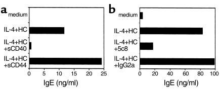
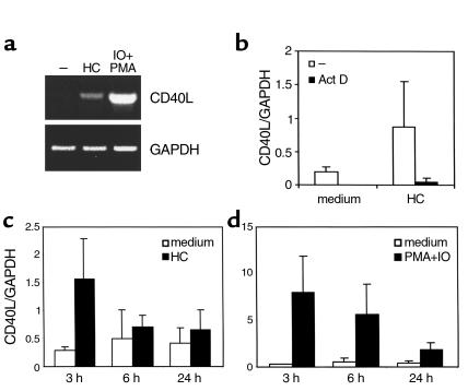
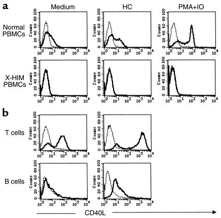
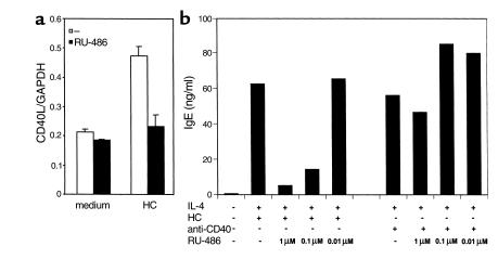
IL-4 and CD40 ligation are essential for IgE synthesis by B cells. We have shown previously that hydrocortisone (HC) induces IgE synthesis in IL-4-stimulated human B cells. In this study we demonstrate that HC fails to induce IgE synthesis in B cells from CD40 ligand-deficient (CD40L-deficient) patients. Disruption of CD40L-CD40 interactions by soluble CD40-Ig fusion protein or anti-CD40L mAb blocked the capacity of HC to induce IgE synthesis in normal B cells. HC upregulated CD40L mRNA expression in PBMCs and surface expression of CD40L in PBMCs as well as in purified populations of T and B cells. Upregulation of CD40L mRNA in PBMCs occurred 3 hours after stimulation with HC and was inhibited by actinomycin D. Upregulation of CD40L mRNA and induction of IgE synthesis by HC were inhibited by the steroid hormone receptor antagonist RU-486. These results indicate that ligand-mediated activation of the glucocorticoid receptor upregulates CD40L expression in human lymphocytes.


Figures

Figure 1
Figure 1
Inhibition of HC+IL-4 induction of IgE synthesis in PBMCs by sCD40-IgG and anti-CD40L mAb. PBMCs from normal donors were stimulated with HC (10–6 M) plus IL-4 (100 U/ml) for 14 days in the absence or presence of (a) sCD40-IgG (5 μg/ml) or control sCD44-IgG (5 μg/ml) and (b) anti-CD40L mAb (5c8, 0.1 μg/ml) or isotype control IgG2a (0.1 μg/ml). Results shown are representative of three experiments.
Figure 2
Figure 2
RT-PCR analysis of HC induction of CD40L expression. (a) PBMCs from a normal donor were stimulated for 3 hours with medium (–), HC (10–6 M), or PMA (20 ng/ml) plus IO (0.5 μM). Equal amounts of total RNA (5 μg) were reverse transcribed, and the cDNA was subjected to RT-PCR using primers that amplify the entire coding region of CD40L cDNA. Final PCR products were analyzed by agarose gel electrophoresis. For equal loading, primers that amplify a 438-bp cDNA fragment of the housekeeping gene GAPDH were used. (b) The effect of actinomycin D (Act D) on upregulation of CD40L mRNA expression by HC are shown. PBMCs were examined for CD40L mRNA expression as in a in the presence or absence of Act D (10 μg/ml). Values represent the mean plus or minus SE of the ratios of the intensity of the bands representing the RT-PCR products of CD40L and GAPDH as assessed by densitometry in three experiments. Kinetics of CD40L mRNA expression in PBMCs stimulated with HC (c) or PMA+IO (d) are shown. Values represent the mean ± SE of the ratios of the intensity of the bands representing the RT-PCR products of CD40L and GAPDH in three experiments.
Figure 3
Figure 3
Upregulation of surface CD40L expression on PBMCs and T and B cells in response to HC stimulation. (a) PBMCs (106/ml) from normal donors or from patients with X-HIM syndrome were cultured for 6 hours with medium, HC (10–6 M), or PMA (20 ng/ml) plus IO (0.5 μM). Cells were then washed and stained with biotin-conjugated anti-CD40L mAb (solid line) or with biotin-conjugated isotype control (dashed line), using the enzymatic amplification method described in Methods, and were analyzed using FACS. Similar results were obtained in two experiments. (b) Purified T or B cells (106/ml) from a normal donor were cultured as in a with either medium or HC. Similar results were obtained in two experiments.
Figure 4
Figure 4
RU-486 inhibits upregulation of CD40L mRNA and induction of IgE synthesis. (a) Effect of RU-486 on CD40L mRNA expression is shown. PBMCs were left unstimulated or were stimulated with HC (10–6 M) for 3 hours in the presence or absence of RU-486 (10–6 M). Values represent the mean plus or minus SE of the ratio of the intensity of the bands representing the RT-PCR products of CD40L and GAPDH in three experiments. (b) Effect of RU-486 on IgE synthesis. PBMCs were stimulated as described in Table 1 in the presence or absence of various concentrations of RU-486. Values represent the mean of net IgE synthesis measured in duplicate-culture supernatants at day 14.

Comment on

  • Corticosteroids, IgE, and atopy.
    Barnes PJ. Barnes PJ. J Clin Invest. 2001 Feb;107(3):265-6. doi: 10.1172/JCI12157. J Clin Invest. 2001. PMID: 11160147 Free PMC article. No abstract available.

References

    1. Vercelli D, Geha RS. Regulation of isotype switching. Curr Opin Immunol. 1992; 4:794–799. - PubMed
    1. Zurawski S, et al. The primary binding subunit of the human interleukin-4 receptor is also a component of the interleukin-13 receptor. J Biol Chem. 1995; 270:13869–13878. - PubMed
    1. Defrance T, et al. Interleukin 13 is a B cell stimulating factor. J Exp Med. 1994; 179:135–143. - PMC - PubMed
    1. Vercelli D, Geha RS. Regulation of IgE synthesis: from the membrane to the genes. Springer Semin Immunopathol. 1993; 15:5–16. - PubMed
    1. Shapira S, Vercelli D, Jabara H, Fu S, Geha R. Molecular analysis of the induction of immunoglobulin E synthesis by human B cells by interleukin 4 and engagement of CD40 antigen. J Exp Med. 1992; 175:289–292. - PMC - PubMed

Publication types