

Functional expression of the new gap junction gene connexin47 transcribed in mouse brain and spinal cord neurons
- PMID: 11160382
- PMCID: PMC3671913
- DOI: 10.1523/JNEUROSCI.21-04-01117.2001
Functional expression of the new gap junction gene connexin47 transcribed in mouse brain and spinal cord neurons
Abstract
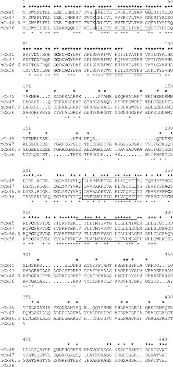
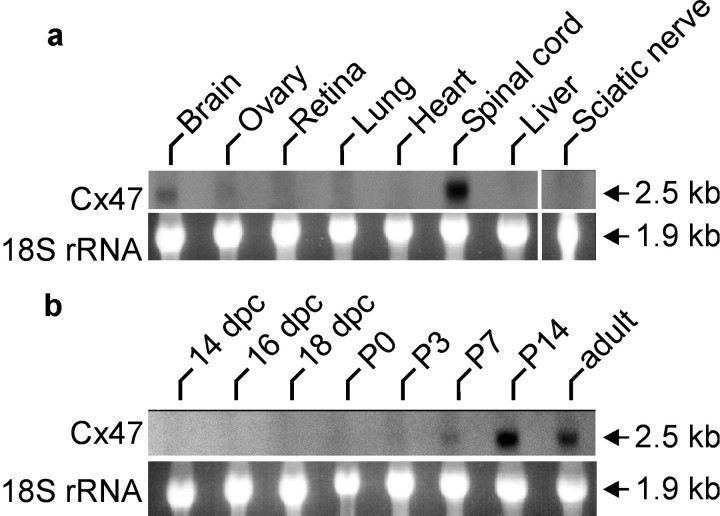
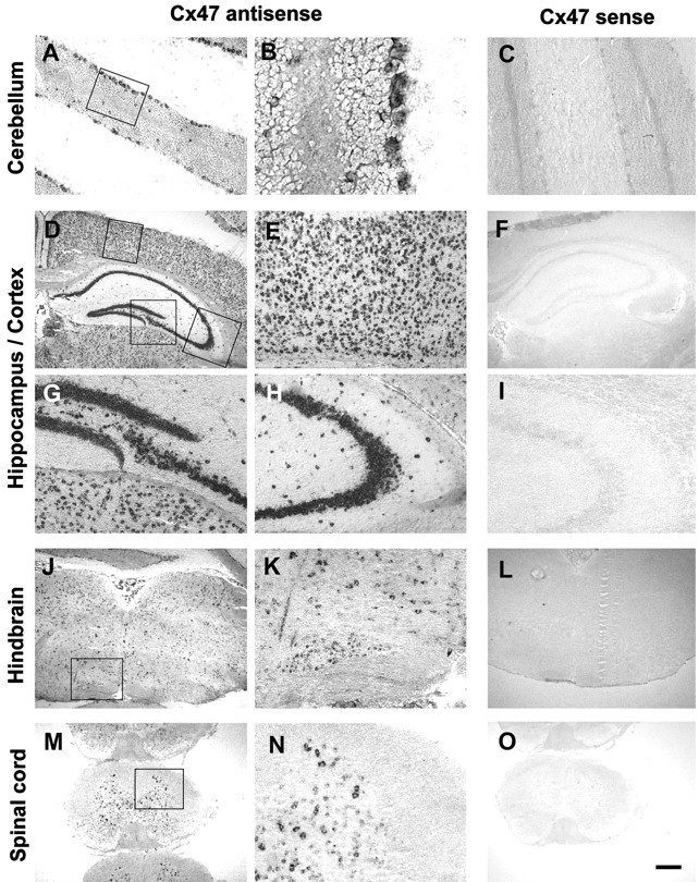
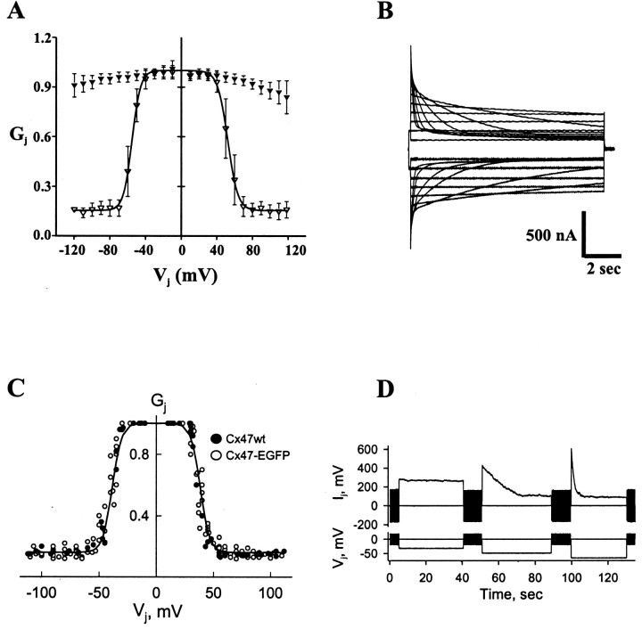
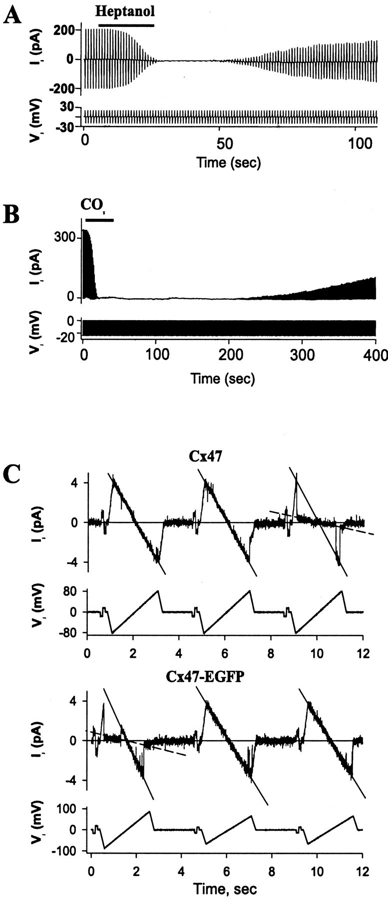
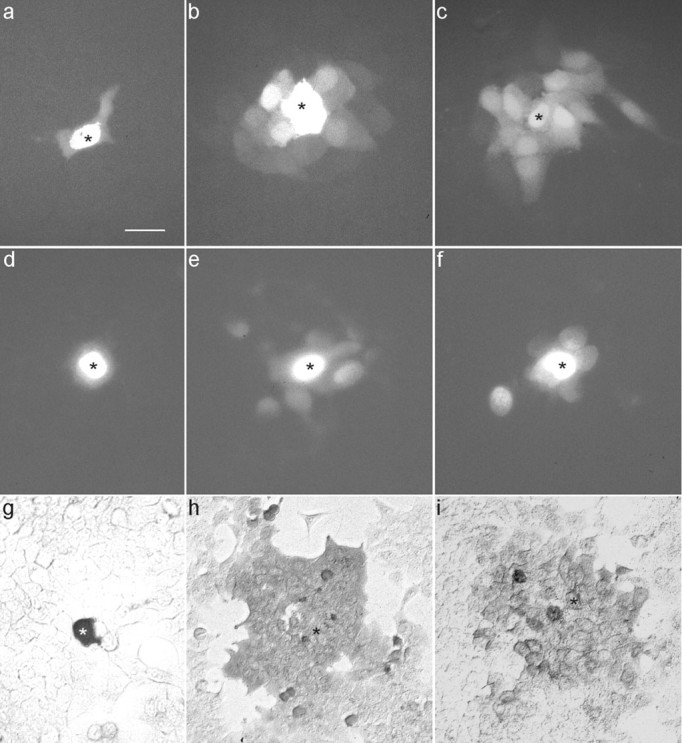
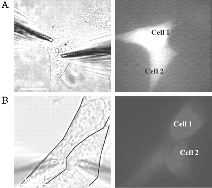
A new mouse gap junction gene that codes for a protein of 46,551 Da has been identified and designated connexin47 (Cx47). It mapped as a single-copy gene to mouse chromosome 11. In human HeLa cells and Xenopus oocytes, expression of mouse Cx47 or a fusion protein of Cx47 and enhanced green fluorescent protein induced intercellular channels that displayed strong sensitivity to transjunctional voltage. Tracer injections in Cx47-transfected HeLa cells revealed intercellular diffusion of neurobiotin, Lucifer yellow, and 4',6-diamidino-2-phenylindole. Recordings of single channels yielded a unitary conductance of 55 pS main state and 8 pS substate. Cx47 mRNA expression was high in spinal cord and brain but was not found in retina, liver, heart, and lung. A low level of Cx47 expression was detected in ovaries. In situ hybridizations demonstrated high expression in alpha motor neurons of the spinal cord, pyramidal cells of the cortex and hippocampus, granular and molecular layers of the dentate gyrus, and Purkinje cells of the cerebellum as well as several nuclei of the brainstem. This expression pattern is distinct from, although partially overlapping with, that of the neuronally expressed connexin36 gene. Thus, electrical synapses in adult mammalian brain are likely to consist of different connexin proteins depending on the neuronal subtype.
Figures
Similar articles
-
Functional expression of the murine connexin 36 gene coding for a neuron-specific gap junctional protein.J Membr Biol. 2000 Aug 1;176(3):249-62. doi: 10.1007/s00232001094. J Membr Biol. 2000. PMID: 10931976 Free PMC article.
-
Molecular and functional diversity of neural connexins in the retina.J Neurosci. 2000 Nov 15;20(22):8331-43. doi: 10.1523/JNEUROSCI.20-22-08331.2000. J Neurosci. 2000. PMID: 11069940 Free PMC article.
-
Functional properties, developmental regulation, and chromosomal localization of murine connexin36, a gap-junctional protein expressed preferentially in retina and brain.J Neurosci Res. 2000 Mar 15;59(6):813-26. doi: 10.1002/(SICI)1097-4547(20000315)59:6<813::AID-JNR14>3.0.CO;2-#. J Neurosci Res. 2000. PMID: 10700019
-
New insights into the expression and function of neural connexins with transgenic mouse mutants.Brain Res Brain Res Rev. 2004 Dec;47(1-3):245-59. doi: 10.1016/j.brainresrev.2004.05.006. Brain Res Brain Res Rev. 2004. PMID: 15572175 Review.
-
Connexin expression systems: to what extent do they reflect the situation in the animal?J Bioenerg Biomembr. 1996 Aug;28(4):319-26. doi: 10.1007/BF02110108. J Bioenerg Biomembr. 1996. PMID: 8844329 Review.
Cited by
-
Reduction of high-frequency network oscillations (ripples) and pathological network discharges in hippocampal slices from connexin 36-deficient mice.J Physiol. 2002 Jun 1;541(Pt 2):521-8. doi: 10.1113/jphysiol.2002.017624. J Physiol. 2002. PMID: 12042356 Free PMC article.
-
Astrocyte and oligodendrocyte connexins of the glial syncytium in relation to astrocyte anatomical domains and spatial buffering.Cell Commun Adhes. 2003 Jul-Dec;10(4-6):401-6. doi: 10.1080/15419060390263191. Cell Commun Adhes. 2003. PMID: 14681048 Free PMC article.
-
Connexin 47 (Cx47)-deficient mice with enhanced green fluorescent protein reporter gene reveal predominant oligodendrocytic expression of Cx47 and display vacuolized myelin in the CNS.J Neurosci. 2003 Jun 1;23(11):4549-59. doi: 10.1523/JNEUROSCI.23-11-04549.2003. J Neurosci. 2003. PMID: 12805295 Free PMC article.
-
Connexins are critical for normal myelination in the CNS.J Neurosci. 2003 Jul 2;23(13):5963-73. doi: 10.1523/JNEUROSCI.23-13-05963.2003. J Neurosci. 2003. PMID: 12843301 Free PMC article.
-
Electrical synapses in mammalian CNS: Past eras, present focus and future directions.Biochim Biophys Acta Biomembr. 2018 Jan;1860(1):102-123. doi: 10.1016/j.bbamem.2017.05.019. Epub 2017 Jun 1. Biochim Biophys Acta Biomembr. 2018. PMID: 28577972 Free PMC article. Review.
References
-
- Adamson MC, Silver J, Kozak CA. The mouse homolog of the Gibbon ape leukemia virus receptor: genetic mapping and a possible receptor function in rodents. Virology. 1991;183:778–781. - PubMed
-
- Al-Ubaidi MR, White TW, Ripps H, Poras I, Avner P, Gomes D, Bruzzone R. Functional properties, developmental regulation, and chromosomal localization of murine connexin36, a gap-junctional protein expressed preferentially in retina and brain. J Neurosci Res. 2000;59:813–826. - PubMed
-
- Belluardo N, Trovato-Salinaro A, Mudo G, Hurd YL, Condorelli DF. Structure, chromosomal localization, and brain expression of human Cx36 gene. J Neurosci Res. 1999;57:740–752. - PubMed
-
- Belluardo N, Mudo G, Trovato-Salinaro A, Le Gurun S, Charollais A, Serre-Beinier V, Amato G, Haefliger J, Meda P, Condorelli DF. Expression of connexin36 in the adult and developing rat brain. Brain Res. 2000;865:121–138. - PubMed
-
- Bennett MVL. Seeing is relieving: electrical synapses between visualized neurons. Nat Neurosci. 2000;3:7–9. - PubMed
Publication types
MeSH terms
Substances
Associated data
- Actions
Grants and funding
LinkOut - more resources
Full Text Sources
Other Literature Sources
Molecular Biology Databases
Miscellaneous