. 2001 Feb 15;21(4):1148-59.
doi: 10.1523/JNEUROSCI.21-04-01148.2001.

Muscarinic regulation of dendritic and axonal outputs of rat thalamic interneurons: a new cellular mechanism for uncoupling distal dendrites

Affiliations

Muscarinic regulation of dendritic and axonal outputs of rat thalamic interneurons: a new cellular mechanism for uncoupling distal dendrites

J Zhu et al. J Neurosci. .

Abstract

Inhibition is crucial for sharpening the sensory information relayed through the thalamus. To understand how the interneuron-mediated inhibition in the thalamus is regulated, we studied the muscarinic effects on interneurons in the lateral posterior nucleus and lateral geniculate nucleus of the thalamus. Here, we report that activation of muscarinic receptors switched the firing pattern in thalamic interneurons from bursting to tonic. Although neuromodulators switch the firing mode in several other types of neurons by altering their membrane potential, we found that activation of muscarinic subtype 2 receptors switched the fire mode in thalamic interneurons by selectively decreasing their input resistance. This is attributable to the muscarinic enhancement of a hyperpolarizing potassium conductance and two depolarizing cation conductances. The decrease in input resistance appeared to electrotonically uncouple the distal dendrites of thalamic interneurons, which effectively changed the inhibition pattern in thalamocortical cells. These results suggest a novel cellular mechanism for the cholinergic transformation of long-range, slow dendrite- and axon-originated inhibition into short-range, fast dendrite-originated inhibition in the thalamus observed in vivo. It is concluded that the electrotonic properties of the dendritic compartments of thalamic interneurons can be dynamically regulated by muscarinic activity.


Figures

Fig. 1.
Fig. 1.
Thalamic interneurons. A,C, Morphology of reconstructed thalamic interneurons in the lateral posterior nucleus (LP) and the dorsal lateral geniculate nucleus (dLGN). Note that these cells were obtained from different slices. B,D, Responses of these interneurons to depolarizing and hyperpolarizing current pulses. E, Plot of the time constants of morphologically identified interneurons (filled circles and filled squaresfor interneurons in LGN and LPN, respectively) and thalamocortical cells (open circles and open squares for thalamocortical cells in LGN and LPN, respectively) against their input resistances. No difference was found in the time constant (τ) or input resistance (Ri) among interneurons (LP: 100.6 ± 7.2 msec, n = 7;LGN: 92.0 ± 3.8 msec, n = 27;t test, p = 0.31 forτ; LPN: 518 ± 37 MΩ,n = 7; LGN: 539 ± 29 MΩ,n = 27; t test,p = 0.72 for Ri) or among thalamocortical cells (LPN: 16.6 ± 1.3 msec, n = 12; LGN: 17.9 ± 1.2 msec, n = 18; t test,p = 0.45 for τ;LPN: 98 ± 9 MΩ, n = 12;LGN: 102 ± 8 MΩ, n = 18;t test, p = 0.77 forRi) in LPN andLGN.
Fig. 2.
Fig. 2.
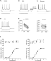
Responses of rat thalamic interneurons to bath application of Mch. A, Responses of a thalamic interneuron to depolarizing and hyperpolarizing current pulses.BD, Bath application of 1 mm Mch (n = 22) or 0.5 mmACh (n = 5) decreased the input resistance of the cell and switched its burst firing pattern to a tonic firing one. Note the reduction of action potential number in the initial burst after application of Mch (Control: 5.2 ± 0.6;Mch: 1.8 ± 0.3; n = 9;p < 0.0001) and large current needed to evoked action potentials during application of agonists. The responses were reversible (not shown for ACh application). E, Input resistance (Ctrl: 602 ± 32 MΩ;Mch: 328 ± 31 MΩ; n = 12;p < 0.0001) and resting membrane potential (Ctrl: −66.1 ± 1.3 mV; Mch: −67.7 ± 1.3 mV; n = 12;p = 0.13) of thalamic interneurons in control or with Mch in bath solution. Large filled circlesrepresent average values (same in the following figures).F, Dose-dependence interneuron responses to bath solution of Mch (left, n = 4) and ACh (right, n = 3). Note the monotonically increasing effect on the input resistance but not on the resting potential. Sigmoid curves in F are the best fitting curves for the average data points from Mch (pKi = 4.50, nH = 0.48) and ACh (pKi = 4.02, nH = 0.45) responses.
Fig. 3.
Fig. 3.
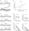
Involvement of muscarinic subtype 2 receptors in Mch-evoked responses in thalamic interneurons. A, Brief bath application of 1 mmMch (4–8 sec) induced a hyperpolarization followed by a depolarization in current-clamp mode or an outward current followed by an inward current in voltage-clamp mode. Note that both hyperpolarization and depolarization were accompanied by a decrease in input resistance (Ctrl: 542 ± 35 MΩ;Hyperpolarization: 397 ± 27 MΩ;p < 0.0001; n = 21;Depolarization: 435 ± 27 MΩ;n = 21; p < 0.0001).B, Plot of peak amplitudes of depolarization (4.2 ± 0.4 mV) against that of hyperpolarization (−6.4 ± 0.6 mV) induced by brief application of Mch (r = 0.51;n = 21; ANOVA, p < 0.05). Linear line is a regression line. C, The muscarinic responses exhibited little desensitization to successive brief applications of 1 mm Mch but were blocked by bath application of 200 nm gallamine. D, Hyperpolarization (Ctrl: 5.6 ± 0.8 mV;Mch: 5.4 ± 0.7 mV; n = 5;p = 0.58) and depolarization (Ctrl: 3.9 ± 0.7 mV; Mch: 3.9 ± 0.4 mV;n = 5; p = 0.87) evoked by the first and second applications of Mch. E, Hyperpolarization (Ctrl: 6.5 ± 0.9 mV;Mch: 0.3 ± 0.2 mV; n = 6;p < 0.001) and depolarization (Ctrl: 4.1 ± 0.9 mV; Mch: 0.2 ± 0.1 mV; n = 6; p < 0.01) in control and with gallamine in bath solution.
Fig. 4.
Fig. 4.
Muscarinic activity enhances a potassium conductance in thalamic interneurons. A, Brief bath application of 1 mm Mch induced a hyperpolarization followed by a depolarization right after the formation of the whole-cell configuration. B, Depolarization was blocked after intracellular loading of 5 mm EGTA for 20–50 min (n = 4). C, I–Vrelationships obtained before (Ctrl) and after application of Mch. Note that current versus voltage (I–V) plots were obtained by ramping membrane potential from −130 or −125 to −55 or −40 mV over a period of 5–10 sec. D, Difference between controlI–V relation and that obtained 5 sec after application of Mch reveals that Mch affected a relatively linear current that reversed at −100.5 ± 1.2 mV (n = 6).
Fig. 5.
Fig. 5.
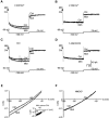
Muscarinic activity enhances two cation conductances in thalamic interneurons. A,C, Stepping voltage to −100 mV before (Ctrl) and 20 sec after application of 1 mm Mch in two interneurons revealed that the slowly activated inward current was augmented during the muscarinic response. The effect was blocked by bath application of 2 mmCs+ (B; n = 7) or intracellular loading of 5 mm EGTA (D;n = 3). Note that the muscarinic activity generated a net outward current when cells were held at a depolarized membrane potential (−0 mV), where Ih was largely inactivated (Zhu et al., 1999b). Calibration in Dapplies also to AC. E,I–V relationships obtained from another interneuron before (Ctrl) and 35–45 sec after application of Mch, whereas 2 mm Cs+ was included in the bath solution. Inset, Subtracting the controlI–V relation from that obtained after application of Mch reveals that Mch induced a linear inward current that reversed at 4.5 ± 3.9 mV (n = 4). F, Substituting extracellular Na+ ions with NMDG+ ions blocked the effect (n= 3).
Fig. 6.
Fig. 6.
Two cation conductances mediate muscarinic depolarization in thalamic interneurons. A, Muscarinic depolarization induced by brief application of 1 mm Mch was partially blocked by bath application of 2 mmCs+ and completely blocked by further substituting extracellular Na+ ions with NMDG+ions. Note that these channel blockers increased the input resistance of the cell. B, Hyperpolarization (Ctrl: 5.8 ± 0.4 mV; Cs+: 7.0 ± 0.3 mV; p < 0.05; n = 6;Cs++NMDG+: 9.0 ± 0.4 mV; n = 6; p < 0.01) and depolarization (Ctrl: 3.7 ± 0.6 mV;Cs+: 1.8 ± 0.3 mV;p < 0.05; n = 6;Cs++NMDG+: −0.4 ± 0.2 mV; n = 6; p< 0.01) in control and with one or two channel blockers.
Fig. 7.
Fig. 7.
Blockade of GABAB inhibition in thalamocortical cells by muscarinic activity. A, Schematic drawing of recording and stimulating electrodes indicates the location of the stimulating electrode, placed within the dendritic tree of the recorded interneuron (∼200 μm away from the soma).B, Direct electric shock evoked a large depolarization and a burst of action potentials in an interneuron in the presence of 5 μm NBQX and 100 μmdl-AP5. Bath application of 1 mm Mch transformed bursting firing into single action potential responses. C, Number of action potentials, averaged over 8–16 trials, in control or with Mch in bath solution (Ctrl: 2.8 ± 0.4; Mch: 0.8 ± 0.1; n = 5; p < 0.01). Circles and squares represent data points obtained from acute and culture slices, respectively (same inF and Fig. 8). D, Schematic drawing of recording and stimulating electrodes indicates the location of the stimulating electrode, placed ∼200–500 μm away from the soma of the recorded thalamocortical cell. E, Direct electric shock evoked a prolonged IPSP in a thalamocortical cell in the presence of 5 μm NBQX and 100 μmdl-AP5. Bath application of 10 μm PTX blocked the early component of the IPSP. The later component of the IPSP was blocked by 1 mm saclofen (n = 3) or 1 mm Mch. F, Amplitude of later IPSPs in control or with Mch in bath solution (Ctrl: 0.51 ± 0.09 mV; Mch: 0.03 ± 0.01 mV;n = 15; p < 0.0001).
Fig. 8.
Fig. 8.
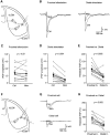
Selective suppression of distal interneuron-mediated GABAA inhibition in thalamocortical cells by muscarinic activity. A, Schematic drawing of recording and stimulating electrodes indicates the locations of two stimulating electrodes, placed ∼250 and 500 μm away from the soma of the recorded thalamocortical cell, respectively. B, Direct electric shock at the proximal and distal locations evoked fast IPSPs in a thalamocortical cell in the presence of 5 μmNBQX, 100 μmdl-AP5, and 1 mmsaclofen. The proximal shock-evoked IPSP was less sensitive to the bath application of 1 mm Mch, compared with the distal shock-evoked one. C, Amplitude of proximal shock-evoked IPSPs in control or with Mch in bath solution (Ctrl: 2.1 ± 0.40 mV; Mch: 1.9 ± 0.4 mV;n = 12; p = 0.52).D, Amplitude of distal shock-evoked IPSPs in control or with Mch in bath solution (Ctrl: 1.3 ± 0.4 mV;Mch: 0.6 ± 0.2 mV; n = 12;p < 0.001). E, Relative amplitude of the proximal shock-evoked and distal shock-evoked IPSPs with Mch in bath solution (Proximal: 91.0 ± 10.0%;Distal: 40.3 ± 8.7%; n = 12;p < 0.005). The values were normalized to control responses. Note that the muscarinic suppression on distal responses was slightly larger in culture slices than in acute slices (culture: 34.6 ± 5.5%; n = 6; acute: 59.6 ± 22.5%; n = 6; t test;p = 0.29). The values were normalized to proximal responses. F, Schematic drawing of recording and stimulating electrodes indicates the simultaneous recordings from two thalamocortical cells, located at ∼250 and 500 μm away from the stimulating electrode. G, Direct electric shock-evoked fast IPSPs in a pair of simultaneously recorded thalamocortical cells in the presence of 5 μm NBQX, 100 μmdl-AP5, and 1 mm saclofen. The IPSP in the proximally located thalamocortical cell was less sensitive to the bath application of 1 mm Mch, compared with the distally located one. H, Relative amplitude of the IPSPs in proximally and distally located thalamocortical cells with Mch in bath solution (Proximal: 89.3 ± 10.3%; Distal: 43.9 ± 8.1%; n = 10; p< 0.005). The values were normalized to control responses. Note that the muscarinic suppression on distal responses was slightly larger in culture slices than in acute slices (culture: 44.5 ± 6.0%;n = 5; acute: 60.1 ± 9.9%;n = 5; t test; p= 0.37). The values were normalized to proximal responses.
Fig. 9.
Fig. 9.
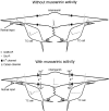
Schematic drawing shows the patterns of interneuron-mediated inhibition in thalamocortical cells with or without muscarinic activity. Note that the sustained muscarinic activity suppresses long-range, slow dendrite- and axon-originated inhibition, but not local, fast dendrite-originated inhibition in thalamocortical cells caused in part by the muscarinic uncoupling of the distal dendrites of thalamic interneurons.

Similar articles

Cited by

References

    1. Ahlsén G, Lindström S, Lo F-S. Inhibition from the brain stem of inhibitory interneurones of the cat's dorsal lateral geniculate nucleus. J Physiol (Lond) 1984;347:593–609. - PMC - PubMed
    1. Ahlsén G, Lindström S, Lo F-S. Interaction between inhibitory pathway to principal cells in the lateral geniculate nucleus of the cat. Exp Brain Res. 1985;58:134–143. - PubMed
    1. Anderson JS, Carandini M, Ferster D. Orientation tuning of input conductance, excitation, and inhibition in cat primary visual cortex. J Neurophysiol. 2000;84:909–926. - PubMed
    1. Bal T, von Krosigk M, McCormick DA. Role of the ferret perigeniculate nucleus in the generation of synchronized oscillations in vitro. J Physiol (Lond) 1995;483:641–663. - PMC - PubMed
    1. Bloomfield SA, Sherman SM. Dendritic current flow in relay cells and interneurons of the cat's lateral geniculate nucleus. Proc Natl Acad Sci USA. 1989;86:3911–3914. - PMC - PubMed

Publication types

MeSH terms

LinkOut - more resources