. 2001 Feb 15;21(4):1302-12.
doi: 10.1523/JNEUROSCI.21-04-01302.2001.

Late oligodendrocyte progenitors coincide with the developmental window of vulnerability for human perinatal white matter injury

Affiliations

Late oligodendrocyte progenitors coincide with the developmental window of vulnerability for human perinatal white matter injury

S A Back et al. J Neurosci. .

Abstract

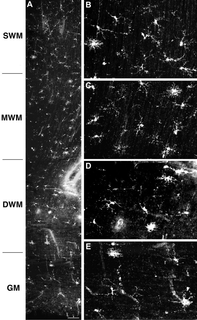
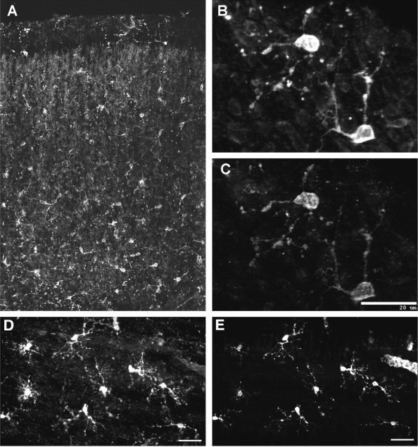
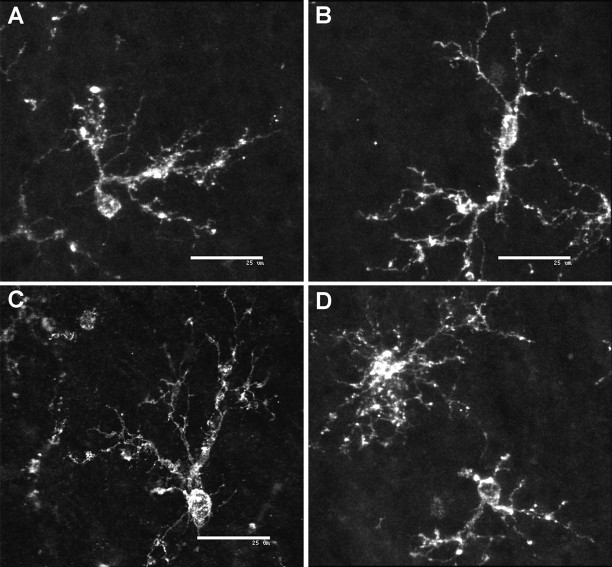
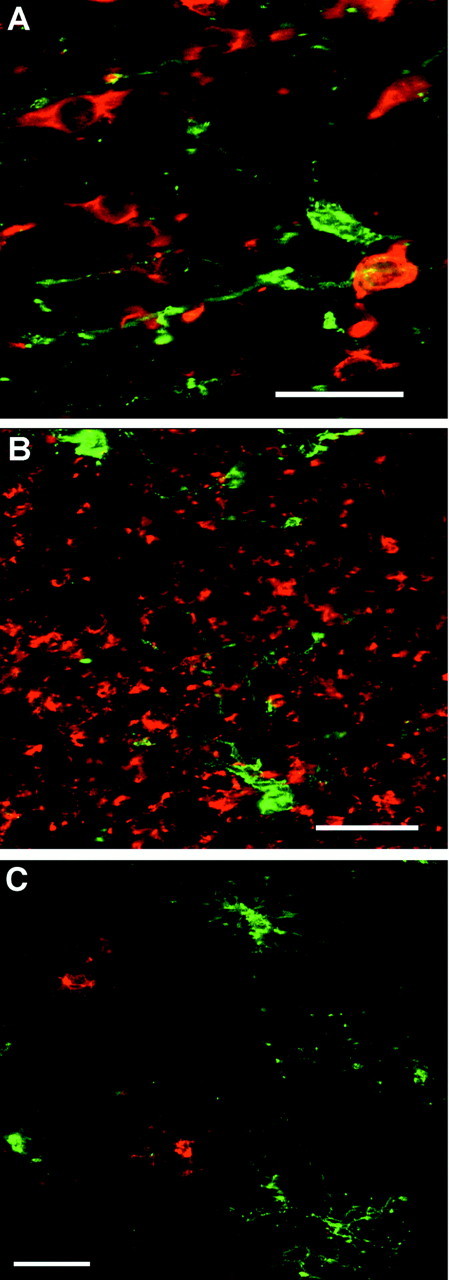
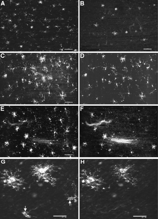
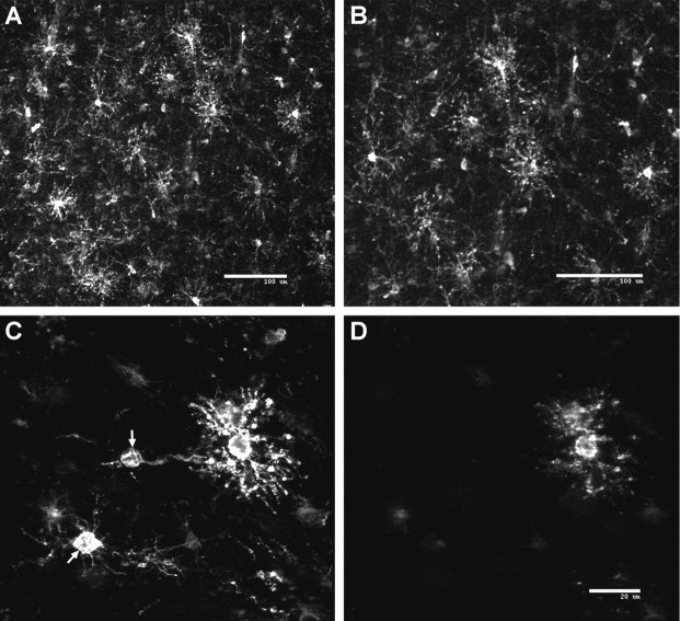
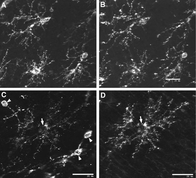
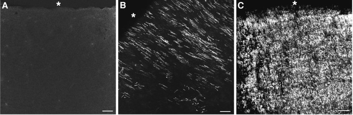
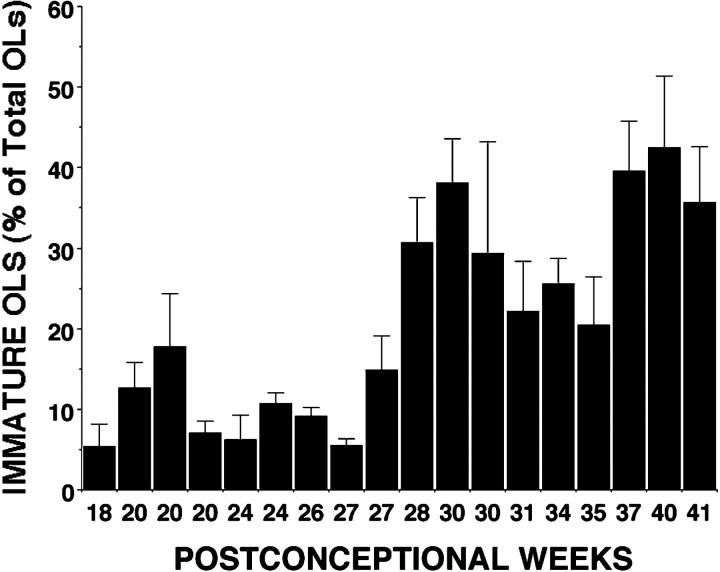
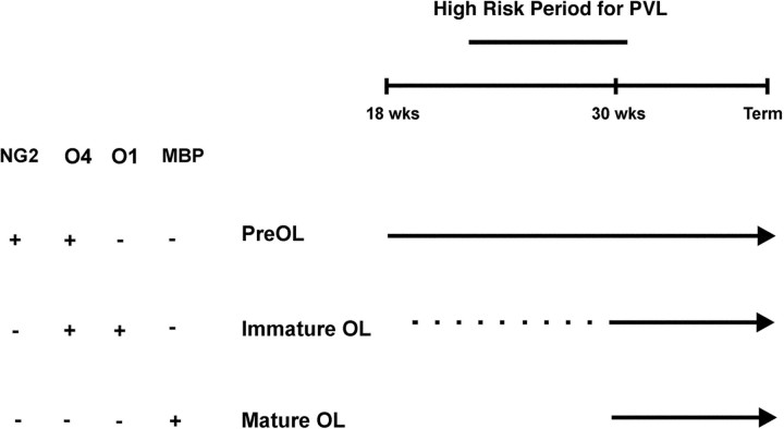
Hypoxic-ischemic injury to the periventricular cerebral white matter [periventricular leukomalacia (PVL)] results in cerebral palsy and is the leading cause of brain injury in premature infants. The principal feature of PVL is a chronic disturbance of myelination and suggests that oligodendrocyte (OL) lineage progression is disrupted by ischemic injury. We determined the OL lineage stages at risk for injury during the developmental window of vulnerability for PVL (23-32 weeks, postconceptional age). In 26 normal control autopsy human brains, OL lineage progression was defined in parietal white matter, a region of predilection for PVL. Three successive OL stages, the late OL progenitor, the immature OL, and the mature OL, were characterized between 18 and 41 weeks with anti-NG2 proteoglycan, O4, O1, and anti-myelin basic protein (anti-MBP) antibodies. NG2+O4+ late OL progenitors were the predominant stage throughout the latter half of gestation. Between 18 and 27 weeks, O4+O1+ immature OLs were a minor population (9.9 +/- 2.1% of total OLs; n = 9). Between 28 and 41 weeks, an increase in immature OLs to 30.9 +/- 2.1% of total OLs (n = 9) was accompanied by a progressive increase in MBP+ myelin sheaths that were restricted to the periventricular white matter. The developmental window of high risk for PVL thus precedes the onset of myelination and identifies the late OL progenitor as the major potential target. Moreover, the decline in incidence of PVL at approximately 32 weeks coincides with the onset of myelination in the periventricular white matter and suggests that the risk for PVL is related to the presence of late OL progenitors in the periventricular white matter.


Figures

Fig. 1.
Fig. 1.
Distribution of O4 mAb-labeled cells in the cerebral white matter at 18 weeks. A, Low-power photomontage showing the regional distribution of cells in the superficial (SWM), mid (MWM), and deep (DWM) cerebral white matter and the germinal matrix (GM). B–E, Detail of the morphology and distribution of cells shown in A in theSWM (B), MWM(C), DWM(D), and GM(E). Scale bar: A, 110 μm.
Fig. 2.
Fig. 2.
A, Low-power photomontage shows the regional distribution of O4 mAb-labeled cells in the cortical mantle from a case at 18 weeks. B–E, Confocal laser digital images demonstrate that OL precursors in the cortical mantle (B, C) and in the cerebral white matter (D, E) labeled for both the O4 antibody (B, D) and NG2 (C, E). Note that the cells in the cortical mantle appear morphologically less mature compared with those in the white matter. Scale bars: B, C, 20 μm; D, E, 25 μm.
Fig. 3.
Fig. 3.
Representative confocal laser microscopic digital three-dimensional reconstructions of NG2-positive cells in the cerebral white matter. Representative examples of the variety of morphological subtypes of pre-OLs identified in the white matter are shown.A, An asymmetric bipolar cell. B, A symmetric bipolar cell. C, An asymmetric multipolar cell. D, Simple and complex multipolar cells. Scale bars: A–D, 25 μm.
Fig. 4.
Fig. 4.
Representative confocal laser microscopic digital images demonstrate the specificity of the O4 antibody for OL precursors. A, B, O4-labeled cells (green) differ in morphology and distribution from neuronal somata (red) in the cortical mantle (A) and from neuronal processes (red) in the superficial white matter (B) that were visualized with anti-β-tubulin isotype III. C, Microglia labeled with the R. communis lectin (red) have a similar morphology but a distinctly different distribution from that of O4-labeled cells (green) in the superficial white matter. Scale bars: A, 20 μm; B, C, 25 μm.
Fig. 5.
Fig. 5.
Pre-OLs are more abundant than immature OLs in human parietal white matter at midgestation. Representative examples of the morphology and distribution of pre-OLs and immature OLs from cases between 18 and 22 weeks of gestation in human parietal white matter are shown. A, B, Cells labeled with O4 (A) were more numerous than those labeled with O1 (B). C, D, The morphology and distribution of cells labeled with the native O4 antibody (C) and a bO4 antibody (D) were quite similar. E, F, Immunofluorescent double labeling with the bO4 (E) and O1 (F) antibodies in the mid cerebral white matter demonstrates that bO4 and O1 labeled a minor population of immature OLs with a complex multipolar morphology. G, H, Confocal laser microscopic digital images are shown of the morphology of complex multipolar immature OLs that labeled with both the bO4 (G) and the O1 (H) antibodies. Note the less differentiated-appearing pre-OLs that labeled with bO4 but not O1 (arrows). Scale bars: A, B, 100 μm; C–F, 60 μm; G, H, 25 μm.
Fig. 6.
Fig. 6.
Pre-OLs persist as the expansion in the immature OL population occurs at ∼30 weeks. Representative confocal laser microscopic digital images from a case at 31 weeks demonstrate the striking change in the morphology of OL precursors that occurs with the expansion of the immature OL population in the parietal white matter.A, B, A low-power image (A) and detail (B) demonstrate the marked increase in complex multipolar cells visualized here in the mid cerebral white matter with the O4 antibody. C, D, Double immunofluorescent-labeling studies with a biotinylated O4 antibody (C) and the O1 antibody (D) revealed that many of the O4-labeled cells were O4+O1− pre-OLs (arrows) that were interspersed among the O4+O1+ immature OLs. Scale bars: A, B, 100 μm; C, D, 20 μm.
Fig. 7.
Fig. 7.
Double immunofluorescent-labeling studies from a case at 32 weeks. Pre-OLs are a morphologically and immunohistochemically distinct population of OL precursors that persist in human cerebral white matter during the expansion in the immature OL population. Pre-OLs were distinguished by diverse morphologies similar to those observed earlier in development. A, B, Representative examples of pre-OLs in the superficial white matter that strongly labeled with O4 (A) and for NG2 (B) are shown. C, Note inC that the NG2-positive pre-OLs (arrowheads) appear morphologically less mature compared with an O1-positive immature OL (D) with a complex multipolar morphology (arrow). Scale bars:A–D, 20 μm.
Fig. 8.
Fig. 8.
The temporal progression of myelinogenesis in human parietal periventricular white matter between 18 and 40 weeks was assessed by immunohistochemical localization of MBP. Representative examples of the distribution of MBP are shown at 20 weeks (A), 30 weeks (B), and 40 weeks (C). The asterisk is adjacent to the ependymal surface of the lateral ventricle. Scale bar:A–C, 100 μm.
Fig. 9.
Fig. 9.
Quantitative analysis of the timing of appearance of pre-OLs and immature OLs in human cerebral white matter between 18 and 41 weeks. Each time point represents an individual case in which the percentage of immature OLs was determined from the total OL somata that labeled with the O1 antibody. Because the O4 antibody was found to label the entire population of OL precursors at all ages examined, total OL precursors were determined from the number of cells that labeled with O4 (see Materials and Methods). Note that O1+ immature OLs are a minor population between 18 and 28 weeks. Approximately 28–30 weeks mark the onset of an expansion in the immature OL population.
Fig. 10.
Fig. 10.
Summary diagram of OL lineage progression in human cerebral white matter during the latter half of gestation. Note that the high-risk period for PVL coincides with the developmental epoch when the white matter is mostly populated by O4+O1− pre-OLs that also label with NG2. O4+O1+ immature OLs are a minor population until ∼30 weeks (dotted line) when they undergo a marked expansion. This expansion in the immature OL population is accompanied by the appearance in the periventricular white matter of MBP+ mature OLs. Hence the decline in incidence of PVL at ∼32 weeks coincides with OL maturation in the periventricular white matter.wks, Weeks.

References

    1. Armstrong RC, Dorn HJ, Kufta CV, Friedman E, Dubois-Dalcq ME. Pre-oligodendrocytes from adult human CNS. J Neurosci. 1992;12:1538–1547. - PMC - PubMed
    1. Back SA, Volpe JJ. Cellular and molecular pathogenesis of periventricular white matter injury. Ment Retard Dev Disabil Res Rev. 1997;3:96–107.
    1. Back SA, Volpe JJ. Approaches to the study of diseases involving oligodendroglial death. In: Koliatsos V, Ratan RR, editors. Cell death and diseases of the nervous system. Humana; Totowa, NJ: 1999. pp. 401–428.
    1. Back SA, Gan X-D, Li Y, Rosenberg PA, Volpe JJ. Maturation-dependent vulnerability of oligodendrocytes to oxidative stress-induced death caused by glutathione depletion. J Neurosci. 1998;18:6241–6253. - PMC - PubMed
    1. Bansal R, Warrington AE, Gard A, Ranscht B, Pfeiffer SE. Multiple and novel specificities of monoclonal antibodies O1, O4, and R-mAb used in the analysis of oligodendrocyte development. J Neurosci Res. 1989;24:548–557. - PubMed

Publication types

MeSH terms

Substances