. 2001 Feb 15;21(4):1393-400.
doi: 10.1523/JNEUROSCI.21-04-01393.2001.

Stimulation of nitric oxide-cGMP pathway excites striatal cholinergic interneurons via protein kinase G activation

Affiliations

Stimulation of nitric oxide-cGMP pathway excites striatal cholinergic interneurons via protein kinase G activation

D Centonze et al. J Neurosci. .

Abstract

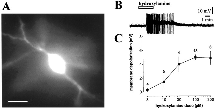
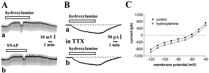
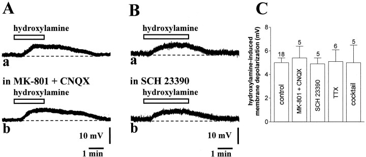
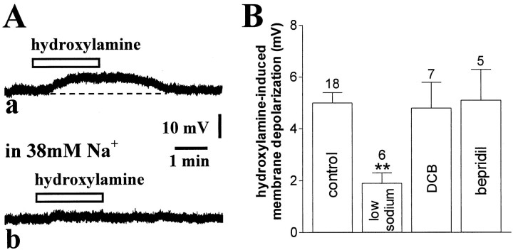
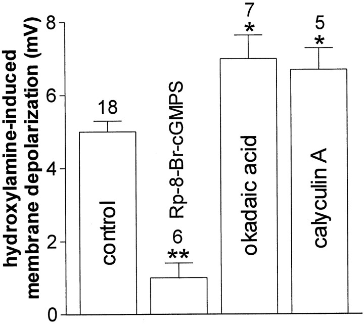
Conflicting data have been collected so far on the action of nitric oxide (NO) on cholinergic interneurons of the striatum. In the present in vitro electrophysiological study, we reported that intracellularly recorded striatal cholinergic interneurons are excited by both hydroxylamine and S-nitroso-N-acetylpenicillamine, two NO donors. This excitation persisted unchanged in the presence of glutamate, dopamine, and substance P receptor antagonists as well as after blockade of tetrodotoxin (TTX)- and calcium channel-sensitive transmitter release, suggesting that NO produces its effects by modulating directly resting ion conductances in the somatodendritic region of striatal cholinergic cells. The depolarizing effect of hydroxylamine was greatly reduced by lowering external concentrations of sodium ions (from 126 to 38 mm) and did not reverse polarity in the voltage range from -120 to -40 mV. The sodium transporter blockers bepridil and 3',4'-dichlorobenzamil were conversely ineffective in preventing NO-induced membrane depolarization. Intracellular cGMP elevation is required for the action of hydroxylamine on striatal cholinergic cells, as demonstrated by the findings that the membrane depolarization produced by this pharmacological agent was prevented by bath and intracellular application of two inhibitors of soluble guanylyl cyclase and was mimicked and occluded by zaprinast, a cGMP phosphodiesterase inhibitor. Finally, intracellular Rp-8-Br-cGMPS, a protein kinase G (PKG) inhibitor, blocked the hydroxylamine-induced membrane depolarization of cholinergic interneurons, whereas both okadaic acid and calyculin A, two protein phosphatase inhibitors, enhanced it, indicating that intracellular PKG and phosphatases oppositely regulate the sensitivity of striatal cholinergic interneurons to NO. The characterization of the cellular mechanisms involved in the regulation of striatal interneuron activity is a key step for the understanding of the role of these cells in striatal microcircuitry.


Figures

Fig. 1.
Fig. 1.
Hydroxylamine depolarizes striatal cholinergic interneurons in a dose-dependent manner. A,Morphological identification of a fura-2-filled striatal cholinergic interneuron. Scale bar, 50 μm. B, Hydroxylamine (100 μm) depolarized a striatal cholinergic interneuron and caused action potential firing. Resting level is −55 mV; full action potential height not captured by pen recorder. C, The graph shows the dose–response curve for the hydroxylamine-induced membrane depolarization of striatal cholinergic interneurons. In this graph and in the following ones, the number of observations is indicated for each experimental condition.
Fig. 2.
Fig. 2.
Electrophysiological characteristics of hydroxylamine- and SNAP-induced excitation of striatal cholinergic interneurons. A, In a current-clamp experiment, hydroxylamine (100 μm) (a) and SNAP (100 μm) (b) depolarized a striatal cholinergic interneuron without affecting the apparent input resistance of the cell. To compare the effects of both compounds on this electrophysiological parameter, membrane potential value was returned to the control level by continuous injection of 100 pA negative current. Resting level was −61 mV. Downward deflections are hyperpolarizing electrotonic potentials evoked by rectangular current pulses (200 pA, 2 sec); their decline after the initial peak reflects the prominent Ih in these cells.B, In another striatal interneuron recorded in the voltage-clamp mode, 100 μm hydroxylamine produced an inward current (a). This effect persisted unchanged in the presence of 1 μm TTX, to block voltage-dependent sodium channels (b). Holding potential was −60 mV. C, Current–voltage relationship of a cholinergic interneuron before (open circles) and during 100 μm hydroxylamine (filled circles). The values were calculated by measuring the steady-state current generated by 3 sec voltage steps of progressively increasing and decreasing amplitude. Holding potential was −60 mV.
Fig. 3.
Fig. 3.
Various receptor antagonists and calcium channel blockers fail to prevent hydroxylamine-induced membrane depolarization of striatal cholinergic interneurons. A, The membrane depolarization produced by 100 μm hydroxylamine (a) was not altered in the presence of 30 μm MK-801 and 10 μm CNQX (7 min) to block both NMDA and non-NMDA glutamate receptors (b). Resting membrane potential was −58 mV. B, Application of the DA D1 receptor antagonist SCH 23390 (10 μm, 7 min) (a) failed to affect the membrane response produced in control medium by 100 μm hydroxylamine (b). Resting membrane potential was −58 mV.C, Summary of pharmacological experiments on 100 μm hydroxylamine-induced membrane depolarization of striatal cholinergic interneurons. Cocktail solution contained: ω-conotoxin GVIA, nifedipine, ω-agatoxin TK, MCPG, and [d--Arg1,d--Pro2,d--Trp7,9,Leu11]-SP. Concentrations were: MK-801 30 μm, CNQX 10 μm, SCH 23390 10 μm, TTX 1 μm, ω-conotoxin GVIA 1 μm, nifedipine 20 μm, ω-agatoxin TK 20 nm, MCPG 300 μm, and [d--Arg1,d--Pro2,d--Trp7,9,Leu11]-SP 10 μm.
Fig. 4.
Fig. 4.
Hydroxylamine-induced membrane depolarization is significantly attenuated by low sodium-containing external solution but not by sodium exchanger blockers. A, In this current-clamp experiment, the membrane depolarization produced by 100 μm hydroxylamine (a) was blocked by 7 min perfusion of the slices with a solution containing 38 mm sodium ions (b). Resting membrane potential was −61 mV. B, Summary of experiments on hydroxylamine (100 μm)-induced membrane depolarization of striatal cholinergic interneurons. Concentration of both DCB and bepridil was 100 μm (**p < 0.01).
Fig. 5.
Fig. 5.
Hydroxylamine-induced membrane depolarization of striatal cholinergic interneurons requires cGMP elevation. A, The pharmacological blockade of sCG by ODQ (5 min, 10 μm) (b) fully prevented the membrane depolarization of a cholinergic interneuron produced by 100 μm hydroxylamine (a). This inhibition was reversible after 15 min wash of this compound (c). Resting membrane potential was −60 mV.B, The cGMP phosphodiesterase inhibitor zaprinast (30 μm) produced a membrane depolarization of another neuron and prevented further depolarization when 100 μmhydroxylamine was added (a). After 10 min wash of both pharmacological agents, the ability of 100 μmhydroxylamine to depolarize the recorded cell was restored and 30 μm zaprinast failed to produce significant depolarization when applied in the presence of this NO donor (b). Resting membrane potential was −62 mV.C, Summary of experiments on hydroxylamine (100 μm)-induced membrane depolarization of striatal cholinergic interneurons. Concentrations were (in μm): extracellular ODQ 10, intraelectrode ODQ 100, and intraelectrode NS 2028 50 (**p < 0.01).
Fig. 6.
Fig. 6.
Role of intracellular PKG and protein phosphatases in the hydroxylamine-induced membrane depolarization of striatal cholinergic cells. The histogram shows the effects of the pharmacological blockade of postsynaptic PKG and protein phosphatases on the membrane depolarization produced by 100 μmhydroxylamine (see Results for details). Concentrations were: Rp-8-Br-cGMPS 1 μm, okadaic acid 30 (n = 3) 100 nm (n = 4), and calyculin A 100 nm (* p < 0.05; **p < 0.01).

References

    1. Aosaki T, Kawaguchi Y. Actions of substance P on rat neostriatal neurons in vitro. J Neurosci. 1996;16:5141–5153. - PMC - PubMed
    1. Aosaki T, Kiuchi K, Kawaguchi Y. Dopamine D1-like receptor activation excites rat striatal large aspiny neurons in vitro. J Neurosci. 1998;18:5180–5190. - PMC - PubMed
    1. Arancio O, Kiebler M, Lee CJ, Lev-Ram V, Tsien RY, Kandel ER, Hawkins RD. Nitric oxide acts directly in the presynaptic neuron to produce long-term potentiation in cultured hippocampal neurons. Cell. 1996;87:1025–1035. - PubMed
    1. Bennett BD, Wilson CJ. Synaptic regulation of action potential timing in neostriatal cholinergic interneurons. J Neurosci. 1998;18:8539–8549. - PMC - PubMed
    1. Black MD, Matthews EK, Humphrey PP. The effects of a photosensitive nitric oxide donor on basal and electrically-stimulated dopamine efflux from the rat striatum in vitro. Neuropharmacology. 1994;33:1357–1365. - PubMed

Publication types

MeSH terms

Grants and funding

LinkOut - more resources