. 2001 Jan 15;21(2):700-12.
doi: 10.1523/JNEUROSCI.21-02-00700.2001.

Cerebellar projections to the prefrontal cortex of the primate

Affiliations

Cerebellar projections to the prefrontal cortex of the primate

F A Middleton et al. J Neurosci. .

Abstract

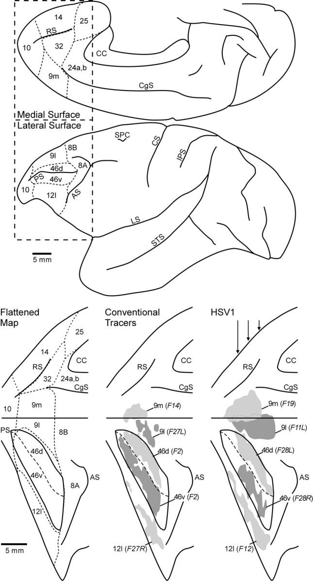
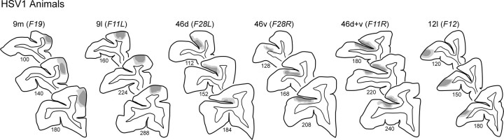
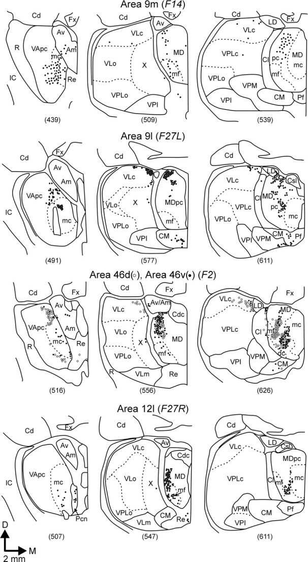
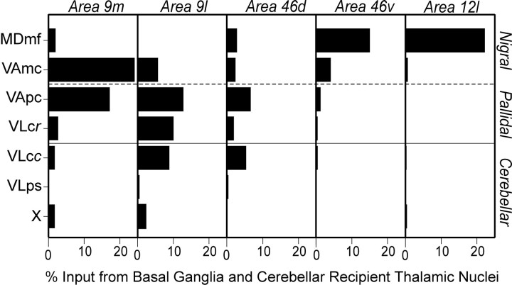
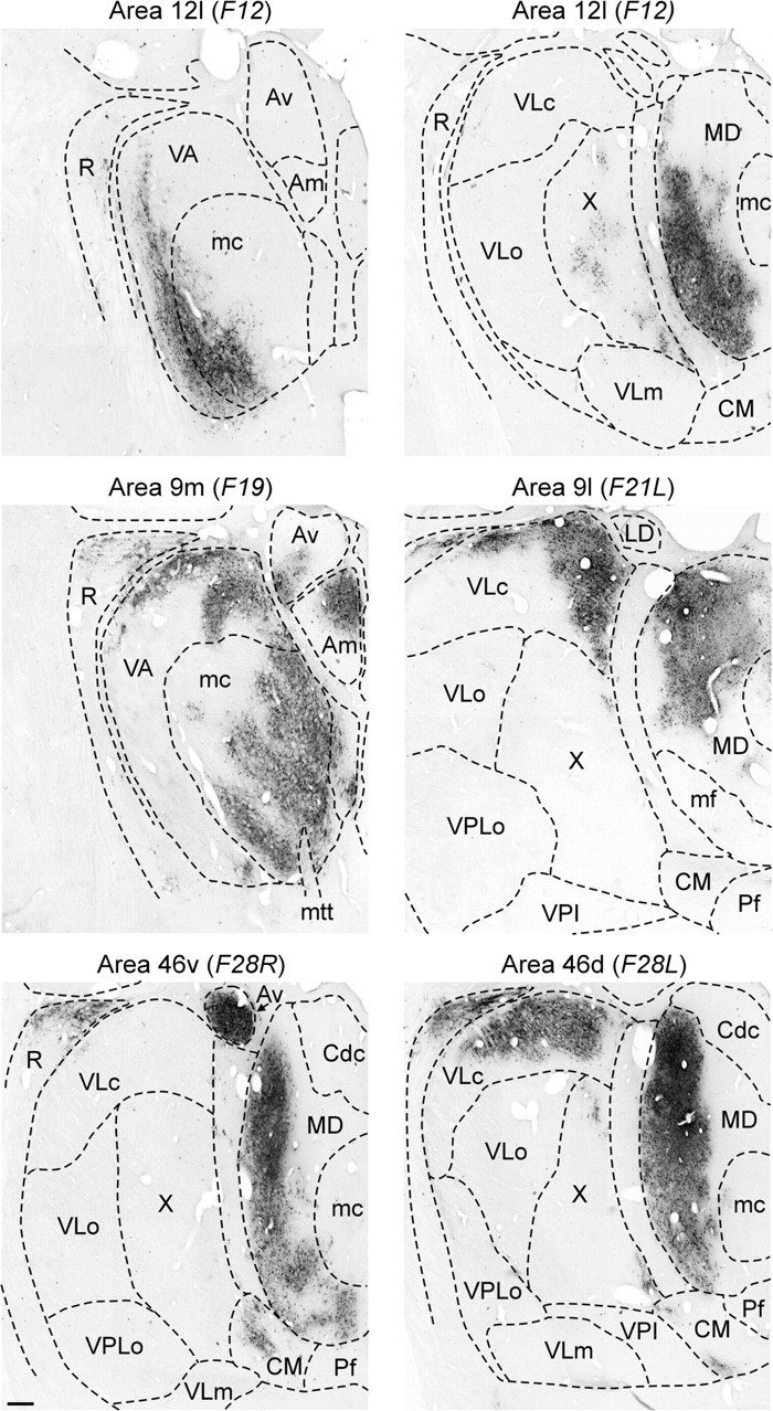
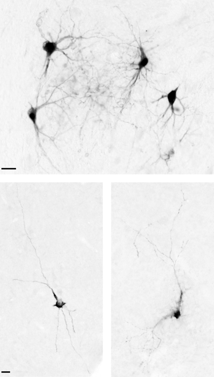
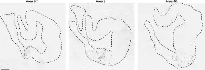
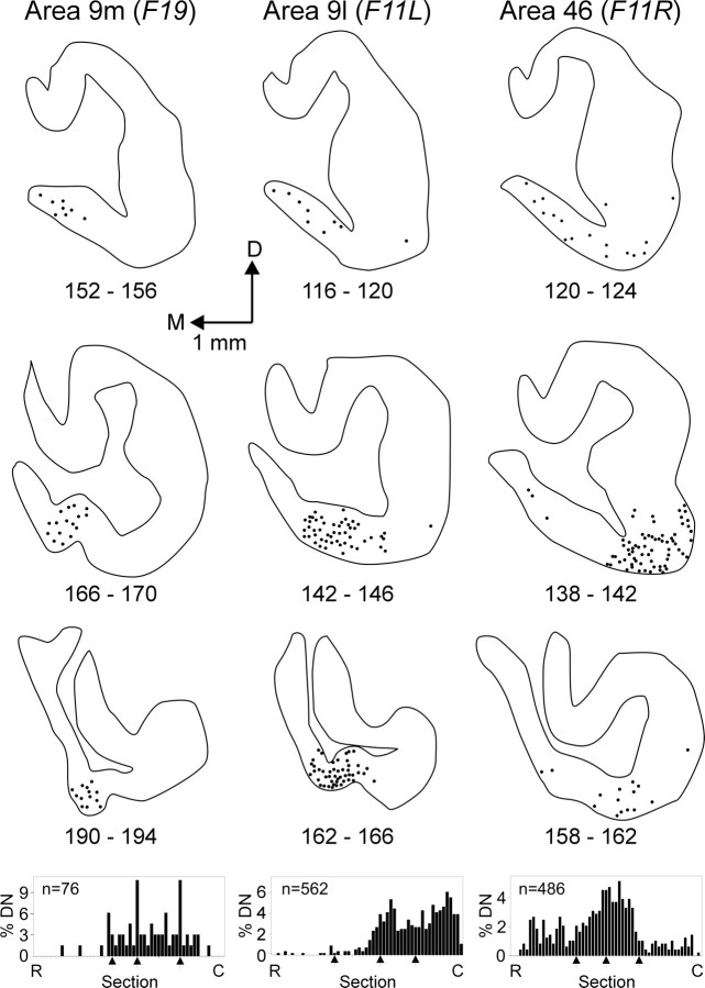
The cerebellum is known to project via the thalamus to multiple motor areas of the cerebral cortex. In this study, we examined the extent and anatomical organization of cerebellar input to multiple regions of prefrontal cortex. We first used conventional retrograde tracers to map the origin of thalamic projections to five prefrontal regions: medial area 9 (9m), lateral area 9 (9l), dorsal area 46 (46d), ventral area 46, and lateral area 12. Only areas 46d, 9m, and 9l received substantial input from thalamic regions included within the zone of termination of cerebellar efferents. This suggested that these cortical areas were the target of cerebellar output. We tested this possibility using retrograde transneuronal transport of the McIntyre-B strain of herpes simplex virus type 1 from areas of prefrontal cortex. Neurons labeled by retrograde transneuronal transport of virus were found in the dentate nucleus only after injections into areas 46d, 9m, and 9l. The precise location of labeled neurons in the dentate varied with the prefrontal area injected. In addition, the dentate neurons labeled after virus injections into prefrontal areas were located in regions spatially separate from those labeled after virus injections into motor areas of the cerebral cortex. Our observations indicate that the cerebellum influences several areas of prefrontal cortex via the thalamus. Furthermore, separate output channels exist in the dentate to influence motor and cognitive operations. These results provide an anatomical substrate for the cerebellum to be involved in cognitive functions such as planning, working memory, and rule-based learning.


Figures

Fig. 1.
Fig. 1.
Location of HSV1 and conventional tracer injections. Top, The lateral surface and medial wall of the cerebral cortex of a cebus monkey. Bottom, The lateral surface and medial wall of the frontal lobe (boxed-in area above) aligned on the midline between the two and both banks of the principal sulcus unfolded.Left, A flattened map of the cytoarchitectonic regions of the prefrontal cortex according to the criteria of Walker (1940) andBarbas and Pandya (1989). Dashed lines indicate the approximate location of the borders between these regions.Middle, The reconstructed injection sites for different conventional tracer experiments. Right, Selected HSV1 injection sites. Shading is used to indicate the combined zones I and II of each injection site, and any regions of overlap are indicated with dotted lines. The approximate locations of the coronal sections through the HSV1 injection sites (see Fig. 2) are indicated with vertical arrowsin the right panel. AS, Arcuate sulcus;CC, corpus callosum; CS, central sulcus;CgS, cingulate sulcus; IPS, intraparietal sulcus; LS, lateral sulcus; PS, principal sulcus; RS, rostral sulcus; SPC, superior precentral sulcus; STS, superior temporal sulcus.
Fig. 2.
Fig. 2.
Cross sections through HSV1 injection sites (see Fig. 1). Low numbers indicate more rostral sections.Dark and light shading are used to indicate the central and peripheral zones, respectively, of the injection site in each section (see Results).
Fig. 3.
Fig. 3.
Thalamic input to the dorsal and lateral prefrontal cortex. Coronal cross sections through representative levels of the thalamus are shown for each conventional tracer experiment. Neurons labeled by retrograde transport from each area are indicated bydots, and nuclear borders are shown bysolid or dotted lines. Thalamic nomenclature and abbreviations are according to Olszewski (1952).D, Dorsal; M, medial; Cd, caudate; c, pars caudalis; dc, pars densocellularis; Fx, fornix; IC, internal capsule; LD, nucleus lateralis dorsalis;mc, pars magnocellularis; mf, pars multiformis; pc, pars parvocellularis; m, pars medialis; R, nucleus reticularis;VPI, nucleus ventralis posterior inferior;VPL, nucleus ventralis posterior lateralis;X, area X.
Fig. 4.
Fig. 4.
Inputs from basal ganglia- and cerebellar-recipient thalamic regions. The percent of total thalamic input to different regions of the prefrontal cortex is shown for those nuclei that are well known targets of basal ganglia and cerebellar projections. Only those basal ganglia- and cerebellar-recipient thalamic nuclei that contained labeling are shown. VLcc, Caudal VLc; VLcr, rostral VLc; ps, pars postrema.
Fig. 5.
Fig. 5.
HSV1-labeled regions of the thalamus. The patterns of HSV1 labeling observed in the thalamus after injections into different regions of the prefrontal cortex are shown at a 5 d survival time. These sections were taken through the most dense regions of thalamic labeling in each case. Conventions are described in Figure 3. Scale bar, 400 μm. mtt, Mamillo thalamic tract.
Fig. 6.
Fig. 6.
HSV1-labeled neurons in the cerebellar output nuclei. Top, HSV1-labeled neurons in the dentate nucleus are shown after injections into area 46 (F1). Bottom left, An example of an HSV1-labeled neuron in the fastigial nucleus is shown after injections of area 9m. Bottom right, An example of an HSV1-labeled neuron in the posterior interpositus nucleus after injection of area 9l. Scale bars, 25 μm.
Fig. 7.
Fig. 7.
Comparison of dentate labeling after HSV1 injections of area 9. Sections at the same level of the dentate are shown after injections of HSV1 into area 9m (left), area 9l (middle), or area 46 (right). Thedashed lines indicate the borders of the dentate nucleus. Note the difference in the location of the labeled neurons within the dentate between cases. Scale bar, 500 μm.
Fig. 8.
Fig. 8.
Origin of dentate projections to areas 9 and 46. Sections through the middle of the dentate nucleus are shown after injections into area 9m (left), area 9l (middle), or area 46 (right). The rostrocaudal distribution of labeled neurons in the dentate for each case is shown at the bottom of the figure, with the locations of the sections shown above indicated by thearrows.DN, Dentate nucleus;R, rostral; C, caudal.

Similar articles

Cited by

References

    1. Akshoomoff NA, Courchesne E. A new role for the cerebellum in cognitive function. Behav Neurosci. 1992;106:731–738. - PubMed
    1. Allen GI, Tsukahara N. Cerebrocerebellar communication systems. Physiol Rev. 1974;54:957–1006. - PubMed
    1. Allen GI, Gilbert PF, Yin TC. Convergence of cerebral inputs onto dentate neurons in monkey. Exp Brain Res. 1978;32:151–170. - PubMed
    1. Andreasen NC, O'Leary DS, Cizaldo T, Arndt S, Rezai K, Boles Ponto LL, Watkins GL, Hichwa RD. Schizophrenia and cognitive dysmetria: a positron-emission tomography study of dysfunctional prefrontal-thalamic-cerebellar circuitry. Proc Natl Acad Sci USA. 1996;93:9985–9990. - PMC - PubMed
    1. Asanuma C, Thach WT, Jones EG. Distribution of cerebellar terminations in the ventral lateral thalamic region of the monkey. Brain Res Rev. 1983;5:237–265. - PubMed

Publication types

Substances

LinkOut - more resources