. 2001 Jan 15;21(2):741-9.
doi: 10.1523/JNEUROSCI.21-02-00741.2001.

Intensity-dependent, rapid activation of presynaptic metabotropic glutamate receptors at a central synapse

Affiliations

Intensity-dependent, rapid activation of presynaptic metabotropic glutamate receptors at a central synapse

G B Awatramani et al. J Neurosci. .

Abstract

Synaptic signals from retinal bipolar cells were monitored by measuring EPSCs in ganglion cells voltage-clamped at -70 mV. Spontaneous EPSCs were strongly suppressed by l-2-amino-4-phosphonobutyrate (AP-4), an agonist at group III metabotropic glutamate receptors (mGluRs). Agonists of group I or II mGluRs were ineffective. AP-4 also suppressed ganglion cell EPSCs evoked by bipolar cell stimulation using potassium puffs, sucrose puffs, or zaps of current (0.5-1 microA). In addition, AP-4 suppressed Off EPSCs evoked by dim-light stimuli. This indicates that group III mGluRs mediate a direct suppression of bipolar cell transmitter release. An mGluR antagonist, (RS)-alpha-cyclopropyl-4-phosphonophenylyglycine (CPPG), blocked the action of AP-4. When bipolar cells were weakly stimulated, AP-4 produced a large suppression of the EPSC, but CPPG alone had little effect. Conversely, when bipolar cells were strongly stimulated, CPPG produced an enhancement of the EPSC, but AP-4 alone had little effect. This indicates that endogenous feedback regulates bipolar cell transmitter release and that the dynamic range of the presynaptic metabotropic autoreceptor is similar to that of the postsynaptic ionotropic receptor. Furthermore, the feedback is rapid and intensity-dependent. Hence, concomitant activation of presynaptic and postsynaptic glutamate receptors shapes the responses of ganglion cells.


Figures

Fig. 1.
Fig. 1.
Effects of mGluR agonists on spontaneous EPSCs in ganglion cells. The ganglion cell was voltage-clamped at −70 mV ( approximate ECl). The spontaneous EPSCs (left) and a histogram and cumulative distribution of the amplitudes (middle) and of the interevent durations (right) of the spontaneous events are shown in the presence of control Ringer's solution (A), the group I mGluR agonist DHPG (B), the group II receptor agonist DCG-IV (C), the group III receptor agonist AP-4 (D), and washout of drugs (E). The asterisk inD (right) indicates that the cumulative distribution was statistically different from control (*p < 0.005 by the Kolmogorov–Smirnov test).
Fig. 2.
Fig. 2.
Group III mGluRs modulate the Off pathway. The light-evoked EPSC in an Off ganglion cell voltage-clamped at −70 mV (ECl) in control Ringer's solution (A), in the presence of AP-4 (B), and when returned to control Ringer's solution (C). The solid barrepresents a 2 sec red-light stimulus.
Fig. 3.
Fig. 3.
Effects of AP-4 on the monosynaptic responses in ganglion cells evoked by artificially stimulating bipolar cells. Responses evoked by electrical pulses (2.5 μA, 1 msec) delivered in the OPL (A), 100 mmK+ puffs (50 msec) in the OPL (B), and 0.5 m sucrose puffs (1 sec) in the IPL (C). Responses are recorded (A–C) in control Ringer's solution (left), in the presence of AP-4 (middle), and after the mGluR agonist was washed out (right). The stimulus artifact preceding the EPSC indicates when the zap was delivered (A). The solid barabove the traces (B,C) represents duration of the stimulus. Neurons were voltage-clamped at −70 mV (ECl).
Fig. 4.
Fig. 4.
Differential effect of mGluR activation on responses evoked by strong and weak stimuli. A, A 5.0 μA current pulse (1 msec) was delivered to the OPL, and the current responses were recorded in a voltage-clamped ganglion cell.B, Weaker current injections of 1.0 μA elicited smaller responses in the ganglion cell. EPSCs in this cell (forA, B) are shown in control Ringer's solution (left), in the presence of AP-4 (middle), and after the agonist has been washed out (right). Neuron was voltage-clamped at −70 mV (ECl).
Fig. 5.
Fig. 5.
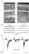
mGluRs inhibit synaptic transmission in the reduced, inner-retinal preparation. A, Photomicrographs of the retinal slice before (i) and after (ii) the outer retina was removed (see Materials and Methods). B, Voltage-clamp recording of the spontaneous activity from a ganglion cell in a reduced slice preparation in control Ringer's solution (i) and in the presence of 2 μm AP-4 (ii). C, Sucrose-evoked (0.5 m, 1 sec) EPSCs recorded from another ganglion cell in a reduced preparation in control Ringer's solution (i), in the presence of 2 μm AP-4 (ii), and after the drug was washed out (iii). The dark barsabovethe traces show when the sucrose was applied. Neurons were voltage-clamped at −70 mV (ECl).
Fig. 6.
Fig. 6.
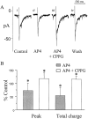
Endogenous activation of mGluRs suppresses bipolar cell output. A, Zap-evoked (5 μA) EPSCs in ganglion cells voltage-clamped at −70 mV in control Ringer's solution (i), in the presence of 2 μm AP-4 (ii), in the presence of 2 μm AP-4 and 200 μm CPPG (iii), and after the drugs were washed out (iv). B, The mean ± SD peak EPSC amplitude and the total EPSC charge caused by the application of AP-4 are represented by gray columns. The CPPG-mediated enhancements of the peak and the total charge are indicated by white columns (*p < 0.05).
Fig. 7.
Fig. 7.
Intensity-dependent modulation of Off responses in ganglion cells. A, Light responses evoked by stimuli of increasing intensity in a ganglion cell voltage-clamped at −70 mV in control Ringer's solution (top) and in the presence of AP-4 (bottom). B, Responses to the same stimulus paradigm recorded in current-clamp mode (zero current), in AP-4 (top), and after the drug was washed out (bottom). C, The mean ± SE of the peak responses from eight cells measured by voltage clamp is plotted as a function of light intensity. The filled squares represent responses measured in control Ringer's solution; the open circles represent responses measured in AP-4. D, Mean AP-4-mediated inhibition as a function of light intensity. The asterisks in Cand D represent mean responses that are statistically different from control (p < 0.01).
Fig. 8.
Fig. 8.
The actions of AP-4 do not depend on the inhibitory circuitry of the retina. All responses were recorded in the presence of 100 μm picrotoxin and 10 μmstrychnine (P + S). A, A series of EPSCs in a ganglion cell evoked by increasing intensities of light stimuli. Responses to the same stimuli were also recorded in the added presence of AP-4 (B) and after AP-4 had been washed out (C). Neurons were voltage-clamped at −70 mV (ECl).
Fig. 9.
Fig. 9.
Bright-light stimuli activate endogenous mGluRs, but weak stimuli do not. For the two types of responses seen in ganglion cells (A, B), responses to dim light are shown in the top row (gray bars) of A and B, whereas responses to bright light (black bars) are shown in thebottom row of A and B. Responses in control (left), in 2 μm AP-4 (middle), and in 2 μm AP-4 plus 200 μm CPPG (right) are illustrated inA and B. The solid gray bars and black bars represent 1 sec (A) and 2 sec (B) of red-light stimuli. Experiments were performed in the presence of 100 μm picrotoxin, 10 μm strychnine, and 500 μm CGP35348. Neurons were voltage-clamped at −70 mV (ECl).

References

    1. Anwyl R. Metabotropic glutamate receptors: electrophysiological properties and role in plasticity. Brain Res Brain Res Rev. 1999;29:83–120. - PubMed
    1. Arkin MS, Miller RF. Subtle actions of 2-amino-4-phosphonobutyrate (APB) on the Off pathway in the mudpuppy retina. Brain Res. 1987;426:142–148. - PubMed
    1. Asztely F, Erdemli G, Kullmann DM. Extrasynaptic glutamate spillover in the hippocampus: dependence on temperature and the role of active glutamate uptake. Neuron. 1997;18:281–293. - PubMed
    1. Awatramani GB, Slaughter MM. Origin of transient and sustained responses in ganglion cells of the retina. J Neurosci. 2000;20:7087–7095. - PMC - PubMed
    1. Brandstätter JH, Koulen P, Kuhn R, van der Putten H, Wässle H. Compartmental localization of a metabotropic glutamate receptor (mGluR7): two different active sites at a retinal synapse. J Neurosci. 1996;16:4749–4756. - PMC - PubMed

Publication types

MeSH terms

LinkOut - more resources