. 2001 Feb;75(4):1689-96.
doi: 10.1128/JVI.75.4.1689-1696.2001.

CD5 is dissociated from the B-cell receptor in B cells from bovine leukemia virus-infected, persistently lymphocytotic cattle: consequences to B-cell receptor-mediated apoptosis

Affiliations

CD5 is dissociated from the B-cell receptor in B cells from bovine leukemia virus-infected, persistently lymphocytotic cattle: consequences to B-cell receptor-mediated apoptosis

G H Cantor et al. J Virol. 2001 Feb.

Abstract

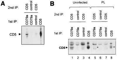
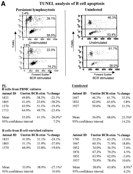
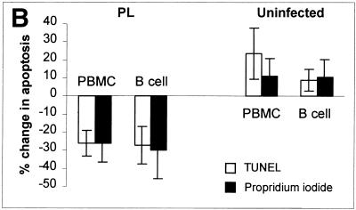
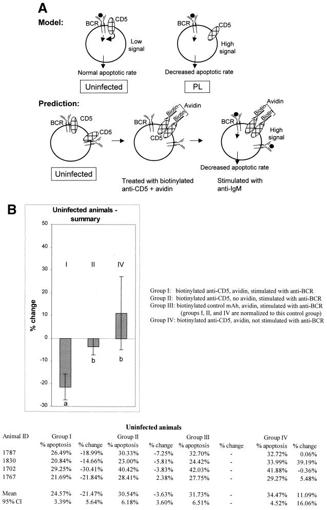
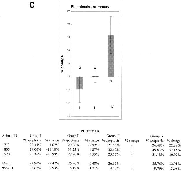
Bovine leukemia virus (BLV), a retrovirus related to human T-cell leukemia virus types 1 and 2, can induce persistent nonneoplastic expansion of the CD5(+) B-cell population, termed persistent lymphocytosis (PL). As in human CD5(+) B cells, we report here that CD5 was physically associated with the B-cell receptor (BCR) in normal bovine CD5(+) B cells. In contrast, in CD5(+) B cells from BLV-infected PL cattle, CD5 was dissociated from the BCR. In B cells from PL cattle, apoptosis decreased when cells were stimulated with antibody to surface immunoglobulin M (sIgM), while in B cells from uninfected cattle, apoptosis increased after sIgM stimulation. The functional significance of the CD5-BCR association was suggested by experimental dissociation of the CD5-BCR interaction by cross-linking of CD5. This caused CD5(+) B cells from uninfected animals to decrease apoptosis when stimulated with anti-sIgM. In contrast, in CD5(+) B cells from PL animals, in which CD5 was already dissociated from the BCR, there was no statistically significant change in apoptosis when CD5 was cross-linked and the cells were stimulated with anti-sIgM. Disruption of CD5-BCR interactions and subsequent decreased apoptosis and increased survival in antigenically stimulated B cells may be a mechanism of BLV-induced PL.


Figures

FIG. 1
FIG. 1
(A) CD5 coimmunoprecipitates with the BCR in uninfected animals. B cells were enriched from PBMCs by T-cell depletion. Lanes 1 to 3, biotin-labeled cells in 1% digitonin lysis buffer were immunoprecipitated (IP) with either MAb to CD79a (the Ig-alpha component of the BCR) or isotype control MAb AV64A. Precipitates were resuspended in 1% NP-40 lysis buffer, and a second immunoprecipitation was performed using MAb to CD5 (MUCIA) or isotype control MAb. As a control, CD5 was immunoprecipitated directly from the PBMC NP-40 lysate using MAb to CD5 (lane 4). (B) CD5 does not coimmunoprecipitate with BCR from BLV-infected PL animals. Methods and design were as in panel A. Despite the presence of a higher proportion of B cells in the PBMCs from the PL animal, CD5 did not coimmunoprecipitate with the BCR (lane 5), although it was seen in coimmunoprecipitations from an uninfected animal (lane 1). A light background band which migrates faster than CD5 is present in lanes 5 to 7. The position of CD5 in the single immunoprecipitation (lane 8) and the predicted position of CD5 in the double immunoprecipitation (lane 5) from PL cells are indicated with asterisks.
FIG. 2
FIG. 2
Apoptosis decreases in BCR-stimulated PL B cells but not in B cells from uninfected animals. The percentage of B cells undergoing apoptosis, as determined by TUNEL analysis, is shown, together with the percent change in apoptosis. Ex vivo PBMCs or B-cell-enriched cell cultures were incubated for 16 h with or without MAb to IgM (1H4; IgG1). Apoptosis was detected using TUNEL (A and B) or propidium iodide staining (B). During FC analyses, B cells were gated for analysis using MAb PIG45A2 (IgG2b) to IgM at 4°C. FC analyses from representative experiments are shown, and the experiments are summarized. In TUNEL analyses, the number of apoptotic cells is defined as the sum of cells in the upper right and left quadrants (A). Significant differences (Student's t test or Mann-Whitney test) are indicated by superscript letters. Means that are significantly different are indicated by different letters, while means that are not statistically different are indicated by the same letter. In panel B, results with TUNEL are compared with those using propidium iodide. The 95% CI are shown. Results with cells from PL animals are significantly different than those with cells from uninfected animals, but are not significantly different based on the technique used to detect apoptosis.
FIG. 2
FIG. 2
Apoptosis decreases in BCR-stimulated PL B cells but not in B cells from uninfected animals. The percentage of B cells undergoing apoptosis, as determined by TUNEL analysis, is shown, together with the percent change in apoptosis. Ex vivo PBMCs or B-cell-enriched cell cultures were incubated for 16 h with or without MAb to IgM (1H4; IgG1). Apoptosis was detected using TUNEL (A and B) or propidium iodide staining (B). During FC analyses, B cells were gated for analysis using MAb PIG45A2 (IgG2b) to IgM at 4°C. FC analyses from representative experiments are shown, and the experiments are summarized. In TUNEL analyses, the number of apoptotic cells is defined as the sum of cells in the upper right and left quadrants (A). Significant differences (Student's t test or Mann-Whitney test) are indicated by superscript letters. Means that are significantly different are indicated by different letters, while means that are not statistically different are indicated by the same letter. In panel B, results with TUNEL are compared with those using propidium iodide. The 95% CI are shown. Results with cells from PL animals are significantly different than those with cells from uninfected animals, but are not significantly different based on the technique used to detect apoptosis.
FIG. 3
FIG. 3
(A) Proposed model and predictions. In B cells from uninfected animals, CD5 is associated with the BCR and downregulates BCR signaling when the BCR is triggered by antigen (indicated by a solid circle). In PL animals, CD5 is dissociated from the BCR and does not downregulate the BCR. The model predicts that in PL animals, in the absence of downregulation by CD5, antigen-triggered BCR signaling will result in decreased apoptosis and increased B-cell longevity. In B cells from uninfected animals, CD5 downregulates antigen-triggered BCR signaling, and apoptosis increases. The model predicts that in B cells from uninfected animals, experimental separation of CD5 from BCR will decrease apoptosis mediated by antigen binding in the same way that apoptosis is decreased in the antigen-triggered PL B cells in which CD5 is already dissociated from the BCR. (B) Cross-linking of CD5 followed by BCR stimulation inhibits apoptosis in CD5+ B cells of uninfected animals. (C) No statistically significant inhibition of apoptosis is seen in CD5+ B cells from PL animals when CD5 is cross-linked followed by BCR stimulation. The percentage of apoptotic cells in each group was compared with group III (non-cross-linked cells stimulated with MAb to BCR) to determine the percent change. Means and 95% CI are shown; significant differences are indicated by superscript letters. In each figure, means that are significantly different are indicated by different letters, while means that are not statistically different are indicated by the same letter.
FIG. 3
FIG. 3
(A) Proposed model and predictions. In B cells from uninfected animals, CD5 is associated with the BCR and downregulates BCR signaling when the BCR is triggered by antigen (indicated by a solid circle). In PL animals, CD5 is dissociated from the BCR and does not downregulate the BCR. The model predicts that in PL animals, in the absence of downregulation by CD5, antigen-triggered BCR signaling will result in decreased apoptosis and increased B-cell longevity. In B cells from uninfected animals, CD5 downregulates antigen-triggered BCR signaling, and apoptosis increases. The model predicts that in B cells from uninfected animals, experimental separation of CD5 from BCR will decrease apoptosis mediated by antigen binding in the same way that apoptosis is decreased in the antigen-triggered PL B cells in which CD5 is already dissociated from the BCR. (B) Cross-linking of CD5 followed by BCR stimulation inhibits apoptosis in CD5+ B cells of uninfected animals. (C) No statistically significant inhibition of apoptosis is seen in CD5+ B cells from PL animals when CD5 is cross-linked followed by BCR stimulation. The percentage of apoptotic cells in each group was compared with group III (non-cross-linked cells stimulated with MAb to BCR) to determine the percent change. Means and 95% CI are shown; significant differences are indicated by superscript letters. In each figure, means that are significantly different are indicated by different letters, while means that are not statistically different are indicated by the same letter.

References

    1. Adam E, Kerkhofs P, Mammerickx M, Kettmann R, Burny A, Droogmans L, Willems L. Involvement of the cyclic AMP-responsive element binding protein in bovine leukemia virus expression in vivo. J Virol. 1994;68:5845–5853. - PMC - PubMed
    1. Berman J J, Moore G W. The role of cell death in the growth of preneoplastic lesions: a Monte Carlo simulation model. Cell Prolif. 1992;25:549–557. - PubMed
    1. Bikah G, Carey J, Ciallella J R, Tarakhovsky A, Bondada S. CD5-mediated negative regulation of antigen receptor-induced growth signals in B-1 B cells. Science. 1996;274:1906–1909. - PubMed
    1. Brooks P A, Cockerell G L, Nyborg J K. Activation of BLV transcription by NF-κB and Tax. Virology. 1998;243:94–98. - PubMed
    1. Burgess K E, Yamamoto M, Prasad K V S, Rudd C E. CD5 acts as a tyrosine kinase substrate within a receptor complex comprising T-cell receptor zeta chain/CD3 and protein-tyrosine kinases p56lck and p59fyn. Proc Natl Acad Sci USA. 1992;89:9311–9315. - PMC - PubMed

Publication types

MeSH terms