. 2001 Feb;75(4):1857-63.
doi: 10.1128/JVI.75.4.1857-1863.2001.

Mechanisms governing expression of the v-FLIP gene of Kaposi's sarcoma-associated herpesvirus

Affiliations

Mechanisms governing expression of the v-FLIP gene of Kaposi's sarcoma-associated herpesvirus

A Grundhoff et al. J Virol. 2001 Feb.

Abstract

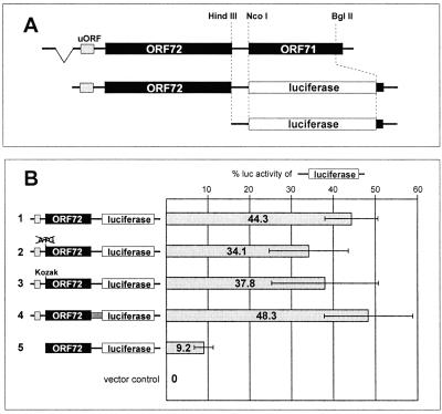
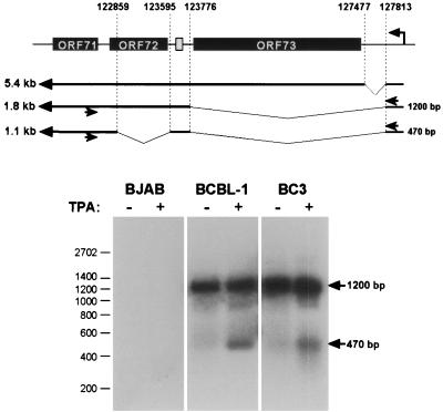
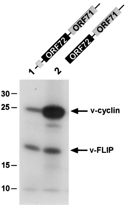
Open reading frame 71 (ORF 71) of Kaposi's sarcoma-associated herpesvirus (KSHV) encodes a death effector domain-containing protein that is homologous to cellular FLIPs (FLICE-inhibitory proteins) and is proposed to inhibit Fas-mediated apoptosis. Transcripts bearing ORF 71 (v-FLIP) sequences are present in all latently infected cells. However, mapping studies reveal these to be bi- or tricistronic mRNAs with ORF 71 located 3' to ORFs 72 (v-cyclin) and 73 (latency-associated nuclear antigen), raising the question of how efficient expression of v-FLIP is achieved. We explored this question by examining the expression of model bicistronic (v-cyclin/LUC) transcripts in which a luciferase (LUC) reporter replaced v-FLIP coding sequences. SLK spindle cells transfected with such constructs efficiently expressed luciferase from the 3' position, and this expression was independent of the expression of the 5' v-cyclin gene. Surprisingly, transcript mapping showed that in these cultures, efficient splicing occurred to remove v-cyclin sequences and generate monocistronic LUC transcripts. Similar splicing events produced monocistronic v-FLIP transcripts in KSHV-infected primary effusion lymphoma cells. However, these RNAs were of low abundance and were inducible by treatment with 12-O-tetradecanoylphorbol-13-acetate. Examination of the more abundant bicistronic latent RNAs revealed the presence of an efficient internal ribosome entry site (IRES) overlapping ORF 72 coding sequences. Thus, two potential mechanisms exist for v-FLIP expression, but the evidence suggests that IRES-mediated internal translational initiation on latent polycistronic mRNAs is the principal source of v-FLIP in latency.


Figures

FIG. 1
FIG. 1
Luciferase expression from bicistronic ORF 72/luciferase reporter constructs. (A) Design of a bicistronic ORF 72/luciferase construct. A genomic fragment containing the second exon of the bicistronic ORF 72/71 mRNA was cloned in the vector pCDNA3, and ORF 71 was replaced by the coding region of the Photinus luciferase. From the resulting construct, pCMV 72/LUC, the region upstream of a HindIII site overlapping the ORF 72 stop codon was excised to generate a monocistronic luciferase construct (pCMV LUC) serving as a positive control. (B) Expression of luciferase from bicistronic reporter constructs in SLK cells. The bicistronic construct pCMV 72/LUC described above is shown in line 1. Additional reporters derived from this construct were modified as follows: the ORF 72 start codon was eliminated (line 2); the nucleotides surrounding the ORF 72 start codon were optimized for translational initiation (12) (line 3); the noncoding segment between ORF 72 and ORF 71 was replaced by an unrelated nucleotide sequence of similar size (line 4); and the region upstream of the ORF 72 start codon was deleted (line 5). The luciferase activity expressed by the monocistronic luciferase construct pCMV LUC described above was set to 100%. Relative luciferase activity of the bicistronic constructs is shown as a percentage of that activity. Each transfection was performed in triplicate per experiment, and the values shown are averaged from three independent experiments.
FIG. 2
FIG. 2
Analysis of luciferase-bearing transcripts in cells transfected with bicistronic reporters. SLK cells were transfected with the bicistronic constructs illustrated in Fig. 1 (lanes 1 to 5), the monocistronic luciferase control pCMV LUC (lane 6), or a vector control (rightmost lane). Total RNA was extracted 36 h posttransfection and analyzed by Northern blotting, using probes specific for luciferase (A) or the 5′-proximal 600 bp of ORF 72 (B). The expected transcript sizes were approximately 3.2 and 3.0 kb for the unspliced bicistronic transcripts in lanes 1 to 4 or lane 5, respectively, and 2.2 kb for the monocistronic luciferase construct in lane 6. The position of the spliced transcripts in lanes 1 to 4 is marked by an arrow in panel A. Bands appear as doublets as a result of usage of two different transcriptional termination sites. The constructs contain the KSHV polyadenylation signal downstream of ORF 71 in addition to the bovine growth hormone polyadenylation signal which is provided by the vector pCDNA3.
FIG. 3
FIG. 3
A 735-nt segment spanning most of the ORF 72 coding region is flanked by functional splice sites. The location of splice sites was determined by RT-PCR amplification and sequencing of splice products as described in Materials and Methods. The genomic sequences flanking the splice donor and acceptor sites are shown in the left and right text boxes, respectively. Within the text boxes, slashes indicate the exact position of the splice donor and acceptor sites, located at nt 123595 and 122859 of the KSHV genome, respectively. Conserved nucleotides (shown in bold) at both sites and a pyrimidine-rich tract (shown underlined) upstream of the acceptor site identify them as consensus splice sites. The start codon of ORF 72 is shown framed within the left text box. The structures of the bicistronic ORF 72/71 and the spliced monocistronic ORF 71 transcripts are illustrated in the lower part of the figure. The locations of the splice sites are indicated by lines emanating from the text boxes. Simple arrows indicate the start codon and the positions of additional AUG codons within the coding region of ORF 72. Arrows marked with asterisks indicate the Kozak start codons (12) which initiate the 5′ uORF and ORF 71. Note that the splice sites are situated to remove the ORF 72 start codon as well as all internal AUG codons, leaving a 66-bp fragment of ORF 72 devoid of potential translation initiation sites.
FIG. 4
FIG. 4
A monocistronic ORF 71 transcript is generated in vivo in PEL cells. Top, location of hybridization sites of RT-PCR primers (indicated by arrows) specific for nt 122566 to 122541 (within ORF 71) and 127871 to 127849 (upstream of ORF 73) of the KSHV genome. The expected size of products amplified from the 1.8-kb bicistronic ORF 72/71 mRNA (1,200 bp) or a putative 1.1-kb monocistronic ORF 71 mRNA (470 bp) is indicated to the right of the transcripts. Bottom, RT-PCR from KSHV-positive and -negative cells using the primers described above. The KSHV-positive PEL cell lines BCBL-1 and BC-3 and the KSHV-negative Burkitt's lymphoma cell line BJAB were treated with TPA to induce lytic replication. Total RNA was isolated from uninduced or induced cells and subjected to RT-PCR as described in Materials and Methods. Aliquots of the samples were analyzed by Southern blotting using a probe encompassing nt 122645 to 122859 of the KSHV genome. The positions of amplification products derived from the bicistronic ORF 72/71 and the monocistronic ORF 71 transcripts are marked by arrows. Sequencing confirmed that the 470-bp product was amplified from a monocistronic RNA resulting from a double splice as illustrated. A product of 290 bp indicative of an ORF 71 transcript generated by a single splice removing nucleotides 122860 to 127812 (see the text) was not detected.
FIG. 5
FIG. 5
An IRES is located upstream of ORF 71. (A) Basic bicistronic Renilla/Photinus (RP) luciferase reporter construct used to detect IRES activity. The construct pCDNA3.1 RP contains the coding region of the Photinus luciferase downstream of the gene for the Renilla luciferase. Expression is driven by the CMV promoter in transfection experiments; a T7 promoter is also available for in vitro transcription (see Fig. 6). Fragments of 232, 474, 658, or 856 bp (the largest fragment contains the complete ORF 72 coding region) upstream of the ORF 71 start codon were inserted in the EcoRI and NcoI sites located in the intercistronic region of the vectors. The authentic ORF 71 start codon was fused to the Photinus luciferase ORF. The 856-bp fragment was also inserted in an antisense orientation (pCMV:RP −856as). The IRES elements from the poliovirus and EMCV served as positive controls. (B) Analysis of transcripts expressed by bicistronic Renilla/Photinus luciferase constructs. Total RNA was isolated from SLK cells 36 h after transfection with the indicated constructs and analyzed by Northern blotting using a probe specific for the Photinus luciferase. Staining of the gel with ethidium bromide showed equal amounts of total RNA loaded in each lane (not shown). (C) IRES activity mediated by the region upstream of ORF 71. SLK cells transfected with the bicistronic constructs were harvested 36 h posttransfection, and Renilla and Photinus luciferase activities were quantitated. The relative luciferase activity (ratio of Photinus to Renilla luciferase activity [P:R]) was calculated and normalized against the activity of the construct pCDNA3.1 RP, whose P:R ratio was set to 1. Each transfection was performed in triplicate.
FIG. 6
FIG. 6
Expression of Renilla/Photinus luciferase in SLK cells transfected with RNA transcribed in vitro from bicistronic RP constructs. RNA was transcribed in vitro as described in Materials and Methods and introduced into 293 cells by electroporation. Aliquots of the cells were harvested at 1.5, 2.5, 3.5, and 4.5 h after electroporation and analyzed for Renilla and Photinus luciferase activity. Shown are the mean values of normalized, relative luciferase activity (see the legend to Fig. 5C) over the total time period of 4.5 h.
FIG. 7
FIG. 7
v-FLIP is translated from a bicistronic ORF 72/71 RNA in vitro. Constructs encoding either the complete second exon of the bicistronic ORF 72/71 RNA (pCMV 72/71; lane 1) or the second exon with a deletion of the 5′ uORF (pCMV 72/71ΔuORF; lane 2) were in vitro transcribed and translated. [35S]methionine-labeled translation products were subsequently analyzed by sodium dodecyl sulfate-polyacrylamide gel electrophoresis and autoradiography. The positions of the v-cyclin and the v-FLIP proteins are indicated by arrows. The identity of the lower band (v-FLIP) was verified by in vitro transcription and translation of constructs in which ORF 71 was deleted or replaced by luciferase (data not shown).

References

    1. Arvanitakis L, Mesri E A, Nador R G, Said J W, Asch A S, Knowles D M, Cesarman E. Establishment and characterization of a primary effusion (body cavity-based) lymphoma cell line (BC-3) harboring Kaposi's sarcoma-associated herpesvirus (KSHV/HHV-8) in the absence of Epstein-Barr virus. Blood. 1996;88:2648–2654. - PubMed
    1. Ballestas M E, Chatis P A, Kaye K M. Efficient persistence of extrachromosomal KSHV DNA mediated by latency-associated nuclear antigen. Science. 1999;284:641–644. - PubMed
    1. Cesarman E, Chang Y, Moore P S, Said J W, Knowles D M. Kaposi's sarcoma-associated herpesvirus-like DNA sequences in AIDS-related body-cavity-based lymphomas. N Engl J Med. 1995;332:1186–1191. - PubMed
    1. Chang Y, Cesarman E, Pessin M S, Lee F, Culpepper J, Knowles D M, Moore P S. Identification of herpesvirus-like DNA sequences in AIDS-associated Kaposi's sarcoma. Science. 1994;266:1865–1869. - PubMed
    1. Chappell S A, Edelman G M, Mauro V P. A 9-nt segment of a cellular mRNA can function as an internal ribosome entry site (IRES) and when present in linked multiple copies greatly enhances IRES activity. Proc Natl Acad Sci USA. 2000;97:1536–1541. - PMC - PubMed

MeSH terms

Associated data