. 2001 Feb;75(4):2024-8.
doi: 10.1128/JVI.75.4.2024-2028.2001.

Does a cdc2 kinase-like recognition motif on the core protein of hepadnaviruses regulate assembly and disintegration of capsids?

Affiliations

Does a cdc2 kinase-like recognition motif on the core protein of hepadnaviruses regulate assembly and disintegration of capsids?

M I Barrasa et al. J Virol. 2001 Feb.

Abstract

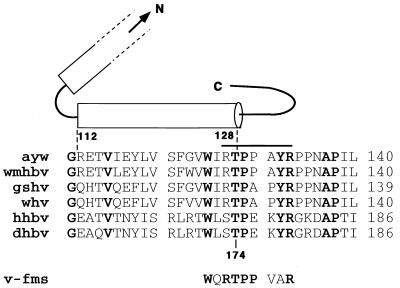
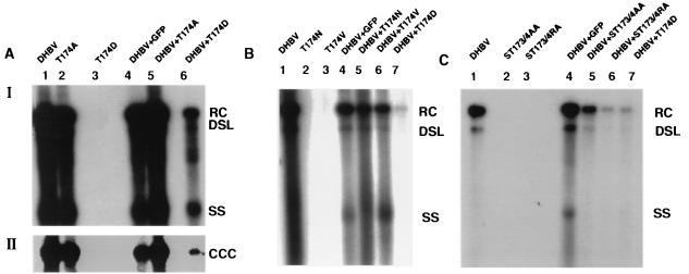
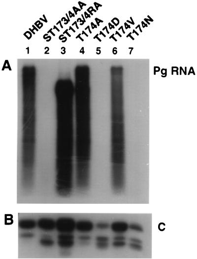
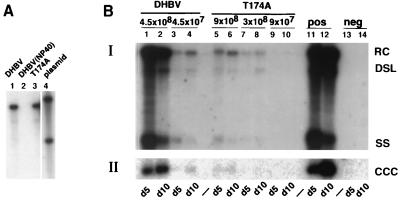
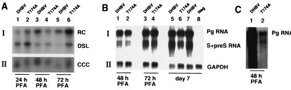
Hepadnaviruses are enveloped viruses, each with a DNA genome packaged in an icosahedral nucleocapsid, which is the site of viral DNA synthesis. In the presence of envelope proteins, DNA-containing nucleocapsids are assembled into virions and secreted, but in the absence of these proteins, nucleocapsids deliver viral DNA into the cell nucleus. Presumably, this step is identical to the delivery of viral DNA during the initiation of an infection. Unfortunately, the mechanisms triggering the disintegration of subviral core particles and delivery of viral DNA into the nucleus are not yet understood. We now report the identification of a sequence motif resembling a serine- or threonine-proline kinase recognition site in the core protein at a location that is required for the assembly of core polypeptides into capsids. Using duck hepatitis B virus, we demonstrated that mutations at this sequence motif can have profound consequences for RNA packaging, DNA replication, and core protein stability. Furthermore, we found a mutant with a conditional phenotype that depended on the cell type used for virus replication. Our results support the hypothesis predicting that this motif plays a role in assembly and disassembly of viral capsids.


Figures

FIG. 1
FIG. 1
Conservation of the threonine-proline kinase motif on hepadnavirus capsid proteins. The figure shows the predicted structure of the HBV capsid protein as described by Bottcher et al. (2). The cylinders represent the complete carboxy-terminal α-helix and a portion of the penultimate α-helix. The bar depicts the segment which is exposed on the surface of core particles, as determined by Pushko et al. (16). The numbering of the amino acids refers to the core sequences of HBV (top) and DHBV (bottom). Aligned were the sequences of HBV (ayw), woolly monkey HBV (wmhbv), ground squirrel and woodchuck hepatitis viruses (gshv and whv), heron virus (hhbv), and DHBV (dhbv). The proline kinase recognition site present on the oncogene v-fms is also shown. Conserved residues are shown in boldface. C, carboxy terminal; N, amino terminal.
FIG. 2
FIG. 2
Viral DNA replication in transfected LMH cells. The figure shows Southern blots of viral core DNA (panels A I, B, and C) and cccDNA (panel AII) extracted from LMH cells (7) transfected with equal amounts of the indicated plasmids. The cells were maintained in Dulbecco's modified Eagle medium–F-12 medium supplemented with 10% fetal bovine serum, kanamycin (100 μg/ml), penicillin (50 U/ml), and streptomycin (50 μg/ml) and transfected with plasmid DNA using a calcium phosphate cell transfection kit (CalPhos mammalian transfection kit; Clontech, Palo Alto, Calif.). Plasmid DHBV directs expression of the DHBV pg from the cytomegalovirus immediate-early promoter (22). Plasmid T174A contains a dA-to-dG substitution at nucleotide 145 (the nomenclature is according to Mandart et al. [12]). Plasmid T174D contains a dAC-to-dGA substitution at nucleotides 145 and 146. Plasmid T174V contains a dAC-to-dGT substitution at nucleotides 145 and 146. Plasmid T174N contains a dC-to-dA substitution at nucleotide 146. Plasmid ST173/4AA contains a dT-to-dG substitution at nucleotide 142 and a dA-to-G substitution at nucleotide 145. Plasmid ST173/4RA contains a dTC-to-dCG substitution at nucleotides 142 and 143 and a dA-to-dG substitution at nucleotide 145. All mutations were verified by nucleotide sequence analysis. RC, relaxed circular, DSL, double-stranded linear; SS; single stranded; CCC, covalently closed circular.
FIG. 3
FIG. 3
Accumulation of packaged pg RNA and core protein in transfected LMH cells. (A) Northern blot analysis of packaged RNA from LMH cells transfected with the indicated plasmids. Encapsidated RNA was extracted as described by Schultz et al. (17). The blot was hybridized with radiolabeled DHBV RNA. (B) Western blot analysis of a total extract from LMH cells transfected with the indicated plasmids. Cells (35-mm-diameter dishes) were lysed in 250 μl of lysis buffer (50 mM Tris-HCl [pH 8], 1% sodium dodecyl sulfate, 1 mM phenylmethylsulfonyl fluoride, 2 μg of leupeptin per ml, and 0.7 μg of pepstatin per ml). Three microliters of extract was analyzed by Western blot using antibodies obtained from a rabbit immunized with DHBV core protein. C, core protein; pg RNA, pregenomic RNA.
FIG. 4
FIG. 4
Infection of PDHs. (A) Southern blot of DNA extracted from enveloped virions present in samples of transfected LMH cells used for the infection of PDHs. A plasmid standard (lane 4) was used to determine the amount of DNA present in lanes 1 and 3. The concentrated culture supernatants were protease treated and incubated with DNase I to remove contaminating core particles, as described by Yu and Summers (27). A sample containing wild-type virus was incubated with NP-40 as a control to demonstrate the efficacy of the protease to digest nonenveloped core particles (lane 2). (B) Southern blot from core (BI) and cccDNA (BII) extracted from PDHs infected with the indicated virus samples. Cells were harvested 5 (d5) and 10 (d10) days postinfection. RC, relaxed circular; DSL, double-stranded linear; SS, single stranded; CCC, covalently closed circular.
FIG. 5
FIG. 5
Formation of cccDNA from virion DNA, viral RNA expression, and packaged RNA in PDHs. (A) Southern blot of core DNA (input virus) (panel AI) and cccDNA (panel AII) extracted from PDHs infected with the indicated virus samples in the presence of PFA. (B) Northern blot analysis of poly(A)-enriched RNA isolated from PDHs infected with the indicated virus samples. The blot was first hybridized with radiolabeled DHBV DNA (panel BI) and subsequently with a probe corresponding to the glyceraldehyde-3-phosphate dehydrogenase (GAPDH) gene (panel BII). Plates were maintained in the presence of PFA and collected 48 and 72 h p.i. or on day 7 p.i. PDHs were infected with 5 × 10⁷ virions per 60-mm culture dish for DHBV and 4 × 10⁷ virions for T174A (panels A and B). (C) Northern blot analysis of packaged RNA from PDHs infected with the indicated virus samples at 3.5 × 10⁸ virions per dish. Encapsidated RNA was extracted as described by Schultz et al. (17). The blot was hybridized with radiolabeled DHBV RNA.

Similar articles

Cited by

References

    1. Beames B, Lanford R E. Insertions within the hepatitis B virus capsid protein influence capsid formation and RNA encapsidation. J Virol. 1995;69:6833–6838. - PMC - PubMed
    1. Bottcher B, Wynne S A, Crowther R A. Determination of the fold of the core protein of hepatitis B virus by electron cryomicroscopy. Nature. 1997;386:88–91. - PubMed
    1. Braaten D, Franke E K, Luban J. Cyclophilin A is required for an early step in the life cycle of human immunodeficiency virus type 1 before the initiation of reverse transcription. J Virol. 1996;70:3551–3560. - PMC - PubMed
    1. Conte D, Jr, Barber E, Banerjee M, Garfinkel D J, Curcio M J. Posttranslational regulation of Ty1 retrotransposition by mitogen-activated protein kinase Fus3. Mol Cell Biol. 1998;18:2502–2513. . (Erratum, 18:5620.) - PMC - PubMed
    1. Conway J F, Cheng N, Zlotnick A, Wingfield P T, Stahl S J, Steven A C. Visualization of a 4-helix bundle in the hepatitis B virus capsid by cryo-electron microscopy. Nature. 1997;386:91–94. - PubMed

Publication types

MeSH terms

LinkOut - more resources