. 2001 Mar;75(5):2130-41.
doi: 10.1128/JVI.75.5.2130-2141.2001.

Antigenicity and immunogenicity of novel chimeric hepatitis B surface antigen particles with exposed hepatitis C virus epitopes

Affiliations

Antigenicity and immunogenicity of novel chimeric hepatitis B surface antigen particles with exposed hepatitis C virus epitopes

H J Netter et al. J Virol. 2001 Mar.

Abstract

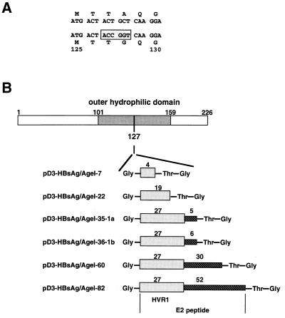
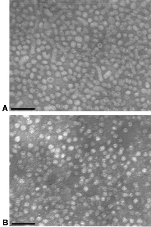
The small envelope protein of hepatitis B virus (HBsAg-S) can self-assemble into highly organized virus like particles (VLPs) and induce an effective immune response. In this study, a restriction enzyme site was engineered into the cDNA of HBsAg-S at a position corresponding to the exposed site within the hydrophilic a determinant region (amino acid [aa] 127-128) to create a novel HBsAg vaccine vector allowing surface orientation of the inserted sequence. We inserted sequences of various lengths from hypervariable region 1 (HVR1) of the hepatitis C virus (HCV) E2 protein containing immunodominant epitopes and demonstrated secretion of the recombinant HBsAg VLPs from transfected mammalian cells. A number of different recombinant proteins were synthesized, and HBsAg VLPs containing inserts up to 36 aa were secreted with an efficiency similar to that of wild-type HBsAg. The HVR1 region exposed on the particles retained an antigenic structure similar to that recognized immunologically during natural infection. VLPs containing epitopes from either HCV-1a or -1b strains were produced that induced strain-specific antibody responses in immunized mice. Injection of a combination of these VLPs induced antibodies against both HVR1 epitopes that resulted in higher titers than were achieved by vaccination with the individual VLPs, suggesting a synergistic effect. This may lead to the development of recombinant particles which are able to induce a broad anti-HCV immune response against the HCV quasispecies or other quasispecies-like infectious agents.


Figures

FIG. 1
FIG. 1
Illustration of the strategy used to insert the HCV HVR1 peptide into the hydrophilic loop of HBsAg-S. (A) Part of the HBsAg-S nucleotide and corresponding amino acid sequence before and after introduction of the AgeI cloning site. The numbers indicate the amino acid position within HBsAg-S. The nucleotide sequence within the rectangle represents the AgeI site. The introduction of this restriction enzyme site leads to an alanine-to-glycine change at position 128. (B) Constructs derived by inclusion of HCV sequences into the AgeI cloning site of the modified HBsAg-S DNA sequence. The first amino acid is glycine, which is not part of the sequence of the HCV E2 protein; the last two amino acids (threonine and glycine) are encoded by the AgeI nucleotide sequence downstream of the HCV E2 insert. The shaded rectangle indicates the HVR1 region of E2, and the hatched rectangle represents the E2 sequence downstream of the HVR1 region. The numbers above the shaded and hatched rectangles indicate the number of encoded amino acids of the corresponding HVR1 region and the downstream E2 region. The HBsAg-S sequence between aa 101 and 159 represents the outer hydrophilic domain.
FIG. 2
FIG. 2
Detection of recombinant HBsAg in cell culture fluid. HuH-7 cells were cotransfected with plasmids encoding one of the HBsAg proteins, a plasmid encoding L-HDAg (see Fig. 3), and pSEAP. Supernatants were harvested, and HBsAg was measured by a chemiluminescence assay. The light counts were normalized by an SEAP assay.
FIG. 3
FIG. 3
Detection of L-HDAg in the presence of the different recombinant HBsAg proteins. (A) Cell culture supernatant (10 ml) (identical samples as used in Fig. 2) was pelleted through a sucrose cushion, resuspended in sample buffer, and analyzed by an immunoblot specific for HDAg. Expression of L-HDAg in the presence of (lane 1) HBsAg wild type, (lane 2) HBsAg/AgeI, (lane 3) HBsAg/AgeI-7, (lane 4) HBsAg/AgeI-22, (lane 5) HBsAg/AgeI-36-1b, (lane 6) HBsAg/AgeI-35-la, (lane 7) HBsAg/AgeI-60, (lane 8) HBsAg/AgeI-82, (lane 9) no HBsAg, and (lane 10) neither HDAg-L nor HBsAg. (B) Analysis of the corresponding cell pellets for the presence of L-HDAg.
FIG. 4
FIG. 4
Equilibrium density gradient analysis of HBsAg VLPs isolated from cell culture fluid. The VLPs were centrifuged through a 20% sucrose cushion, resuspended in PBS, and then centrifuged to equilibrium on preformed step gradients of CsCl (10 to 40% [wt/wt]). (A) Wild-type (wt) HBsAg particles. (B) Recombinant particles expressing HVR1-1b.
FIG. 5
FIG. 5
Identification of particles by electron microscopy. (A) Particles derived from the serum of a chronic HBV carrier. (B) Recombinant particles derived from the construct pD3-HBsAg/AgeI-36-1b. Bars, 100 nm.
FIG. 6
FIG. 6
Reactivity of recombinant particles with human serum as determined by ELISA. (A) Serum from a patient infected with HCV was tested against peptides representing HVR1-1a and HVR1-1b sequences and an unrelated peptide. (B and C) Two independently performed assays testing the human serum against recombinant (rec.) HBsAg particles containing the HVR1-1a epitope or HVR1-1b epitope, wild-type (wt) HBsAg particles, and a control fraction derived from the cell culture fluid of untransfected HuH-7 cells (mock).
FIG. 7
FIG. 7
Immunogenicity of recombinant VLPs in mice as determined by ELISA. Serum samples were from a mouse immunized with HBsAg/AgeI-35-1a recombinant particles (A) or HBsAg/AgeI-35-1b recombinant particles (B). The mice were immunized three times on days 0, 33, and 47, and serum samples were taken on days 56, 69, 82, and 110 and tested (1:50 and 1:200 dilutions) against the HVR1-1a peptide, the HVR1-1b-specific peptide, and an unrelated peptide. The results show the mean OD of multiple tests and the standard deviation.
FIG. 8
FIG. 8
Induction of antibodies in mice immunized with a combination of VLPs as determined by ELISA. The mice were immunized three times on days 0, 33, and 47, and serum samples were taken on days 56, 69, 82, 110, and 143. The results shown are from a representative mouse immunized with a combination of HBsAg/AgeI-35-1a and HBsAg/AgeI-35-1b recombinant particles. Serum samples (diluted 1:50 and 1:200) were tested against the HVR1-1a peptide, the HVR1-1b-specific peptide, and an unrelated peptide. The results show the mean OD of multiple tests and the standard deviation.
FIG. 9
FIG. 9
Antibody titer against HVR1 epitopes in mouse sera taken at different time points, shown as a function of time. Mice were immunized on days 0, 33, and 47 (indicated by arrows), and the serum was taken on days 0 (prebleed), 47, 56, 69, 82, 110, 143, and 209 (A1, B1 and C), or mice were immunized on days 0 and 15 and serum was taken on days 0 (prebleed), 24, 44, and 57 (A2 and B2). Antibody titers against the HVR1-1a epitope in two mice immunized with HBsAg/Age-35-1a (A1 and A2), against HVR1-1b in one mouse immunized with HBsAg/Age-36-1b (B1 and B2), and in three mice immunized with a combination of particles (C). ○, antibody titer against the HVR1-1a epitope; ●, antibody titer against the HVR1-1b epitope. The titer is given as the arimethic mean, and the bars indicate the range of antibody titers obtained. The values shown in panels A2, B2, and C indicate titers outside the range of the y axis.
FIG. 9
FIG. 9
Antibody titer against HVR1 epitopes in mouse sera taken at different time points, shown as a function of time. Mice were immunized on days 0, 33, and 47 (indicated by arrows), and the serum was taken on days 0 (prebleed), 47, 56, 69, 82, 110, 143, and 209 (A1, B1 and C), or mice were immunized on days 0 and 15 and serum was taken on days 0 (prebleed), 24, 44, and 57 (A2 and B2). Antibody titers against the HVR1-1a epitope in two mice immunized with HBsAg/Age-35-1a (A1 and A2), against HVR1-1b in one mouse immunized with HBsAg/Age-36-1b (B1 and B2), and in three mice immunized with a combination of particles (C). ○, antibody titer against the HVR1-1a epitope; ●, antibody titer against the HVR1-1b epitope. The titer is given as the arimethic mean, and the bars indicate the range of antibody titers obtained. The values shown in panels A2, B2, and C indicate titers outside the range of the y axis.

Similar articles

Cited by

References

    1. Berger J, Hauber J, Hauber R, Geiger R, Cullen B R. Secreted placental alkaline phosphatase: a powerful new quantitative indicator of gene expression in eucaryotic cells. Gene. 1988;66:1–10. - PubMed
    1. Bruss V, Gerhardt E, Vieluf K, Wunderlich G. Functions of the large hepatitis B virus surface protein in viral particle morphogenesis. Intervirology. 1996;39:23–31. - PubMed
    1. Chang F-L, Chen P-J, Tu S-J, Wang C-J, Chen D-S. The large form of hepatitis δ antigen is crucial for assembly of hepatitis δ virus. Proc Natl Acad Sci USA. 1991;88:8490–8494. - PMC - PubMed
    1. Chengalvala M V, Bhat R A, Bhat B M, Vernon S K, Lubeck M D. Enhanced immunogenicity of hepatitis B surface antigen by insertion of a helper T cell epitope from tetanus toxoid. Vaccine. 1999;17:1035–1041. - PubMed
    1. Choo Q L, Kuo G, Ralston A, Weiner A, Chien D, Van Nest G, Han J, Berger K, Thudium K, Kuo C, Kansopon J, McFarland J, Tabrizi A, Ching K, Moss B, Cummins L B, Houghton M, Muchmore E. Vaccination of chimpanzees against infection by the hepatitis C virus. Proc Natl Acad Sci USA. 1994;91:1294–1298. - PMC - PubMed

Publication types

MeSH terms

LinkOut - more resources