. 2001 Jan;12(1):1-12.
doi: 10.1091/mbc.12.1.1.

Serine phosphorylation of focal adhesion kinase in interphase and mitosis: a possible role in modulating binding to p130(Cas)

Affiliations
Free PMC article

Serine phosphorylation of focal adhesion kinase in interphase and mitosis: a possible role in modulating binding to p130(Cas)

A Ma et al. Mol Biol Cell. 2001 Jan.
Free PMC article

Abstract

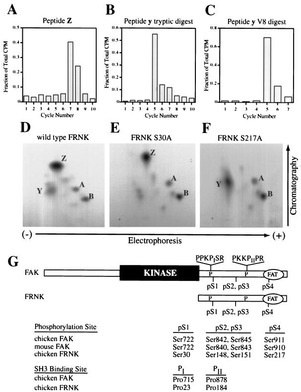
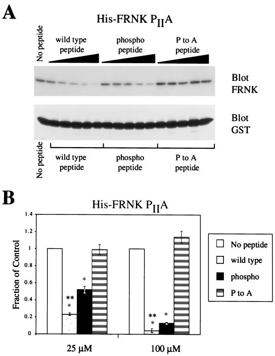
Focal adhesion kinase (FAK) is an important regulator of integrin signaling in adherent cells and accordingly its activity is significantly modulated during mitosis when cells detach from the extracellular matrix. During mitosis, FAK becomes heavily phosphorylated on serine residues concomitant with its inactivation and dephosphorylation on tyrosine. Little is known about the regulation of FAK activity by serine phosphorylation. In this report, we characterize two novel sites of serine phosphorylation within the C-terminal domain of FAK. Phosphorylation-specific antibodies directed to these sites and against two previously characterized sites of serine phosphorylation were used to study the regulated phosphorylation of FAK in unsynchronized and mitotic cells. Among the four major phosphorylation sites, designated pS1-pS4, phosphorylation of pS1 (Ser722) is unchanged in unsynchronized and mitotic cells. In contrast, pS3 and pS4 (Ser843 and Ser910) exhibit increased phosphorylation during mitosis. In vitro peptide binding experiments provide evidence that phosphorylation of pS1 (Ser722) may play a role in modulating FAK binding to the SH3 domain of the adapter protein p130(Cas).


Figures

Figure 1
Figure 1
FRNK, the autonomously expressed carboxyl terminus of FAK, is phosphorylated in vivo on Ser30 (pS1) and Ser217 (pS4). (A–C) Sequential Edman degradation of phosphopeptides collected from in vivo labeling of FRNK. Phosphopeptides were isolated from TLC plates and subjected to sequential Edman degradation as described in MATERIALS AND METHODS. The amount of radioactivity released was determined after each cycle of degradation. Tryptic peptide Z was collected from CE cells overexpressing wild-type FRNK and labeled in vivo with [32P]orthophosphate (A). For peptide y, GST-FRNK was phosphorylated in vitro with protein kinase A and unlabeled ATP, followed by labeling with casein kinase I and [γ-32P]ATP. GST-FRNK was digested with trypsin (B) or V8 (C) before TLC analysis and Edman degradation. Values are expressed as a fraction of total radioactivity recovered after the indicated number of cycles. (D–F) Two-dimensional phosphopeptide mapping of wild-type FRNK (D) or FRNK point mutants at pS1 (E) or pS4 (F) expressed in CE cells labeled in vivo with [32P]orthophosphate. (G) Unified nomenclature for FAK and FRNK serine phosphorylation sites and critical Cas-binding proline residues based on their relative positions in each protein. ‘FAT‘ represents the focal adhesion-targeting sequence at the extreme C termini of FAK and FRNK.
Figure 2
Figure 2
Site-specific recognition of serine-phosphorylated residues in the C terminus of FAK by phospho-specific antibodies raised against each site of phosphorylation. (A) Myc-tagged wild-type FAK or FAK variants harboring mutations at pS2 or pS3 were transfected into HeLa cells. Lysates of transfected cells were immunoblotted with anti-myc (lanes 1-3), anti-pS2 (lanes 4-6), or anti-pS3 (lanes 7-9) to demonstrate that mutation of the immunogenic phosphorylated serine residue abolished epitope recognition by the phospho-specific antibodies. Similarly, in B, FLAG-tagged FAK or FAK variants harboring mutations at pS1 or pS4 were tested with the anti-FLAG (lanes 1-3), anti-pS1 (lanes 4-6), or anti-pS4 (lanes 7-9) antibodies. Endogenous FAK is indicated by the open arrows, ectopically expressed FAK by the closed arrows.
Figure 3
Figure 3
Recognition of FAK by phospho-specific antibodies against serine phosphorylated antigens and specific competition by phosphorylated peptides. (A) Reactivity of the phospho-specific antibodies with immune complexes of endogenous FAK (lanes 3 and 4, 7 and 8, 11 and 12, and 15 and 16) and proteins in lysates (lanes 5 and 6, 9 and 10, 13 and 14, and 17 and 18) of unsynchronized (U) and mitotic (M) HeLa cells. FAK is indicated by the arrow; additional cross-reactive bands are indicated by dots (●). (B) Reactivity of the phospho-specific antibodies in Western blots can be specifically blocked by preincubation with phosphorylated synthetic peptide antigen. Each phospho-specific antibody was preincubated at a working concentration of 2-3 μg/ml in the absence of peptide or with a 20- to 100-fold molar excess of nonphosphorylated or phosphorylated peptide. Nitrocellulose strips containing U and M HeLa cell proteins resolved by SDS-PAGE were blocked and then incubated with the pretreated antibody solutions. Total FAK was detected by immunoblotting with the monoclonal antibody 2A7 (lanes 1 and 2). The band corresponding to FAK is indicated by the open arrow; in the anti-pS2 blot, FAK appears as a weak signal migrating above the strong band at ∼120 kDa.
Figure 4
Figure 4
A synthetic peptide derived from the site I sequence of FAK specifically interacts with the SH3 domain of Cas. GST alone (lanes 1 and 2) or fused to the SH3 domain of Cas (lanes 3-8), cortactin (lanes 9 and 10), or Src (lanes 11 and 12), or the N terminus of paxillin (lanes 13 and 14) was preincubated with increasing amounts of wild-type peptide (EAPPKPSRPGYPSPRSS, 0 to 100 μM), followed by incubation with 500 μg of CE cell lysate. Protein complexes were captured, resolved by SDS-PAGE, and blotted for the presence of FAK (top) and FRNK (bottom) with the polyclonal antiserum BC3.
Figure 5
Figure 5
Analysis of the binding of phosphorylated peptides derived from the site I sequence to the Cas SH3 domain. (A) Representative Western blot of peptide competition assays by using GST-CasSH3 and wild-type His-tagged FRNK. Components were incubated in the presence of increasing amounts (1, 10, 25, 50, or 100 μM) of each peptide indicated. His-FRNK was detected by immunoblotting with the mAb 2A7 followed by detection with 125I anti-mouse IgG and densitometric analysis. (B) Quantitation and graphical representation of PhosphorImager data with wild-type His-FRNK with 25 or 100 μM peptide competitor. Values are expressed as a fraction of a control reaction performed in the absence of peptide (open bar, normalized to 1). P values were calculated using a paired t test. Statistical differences (P < 0.05) between either the wild-type peptide or the phosphopeptide and the control P→A peptide are indicated by an asterisk (∗) above the appropriate bar. Statistical differences between the wild type and phosphopeptides themselves are indicated by a double asterisk (∗∗) above the bar representing the wild-type peptide. Data points represent mean values from three independent experiments; error bars represent SD from the mean.
Figure 6
Figure 6
Effects of serine phosphorylation on FAK peptide binding to the Cas SH3 domain when Cas binding is restricted to site I of His-FRNK. (A) Representative Western blot of peptide competition assays by using GST-Cas SH3 and His-FRNK PIIA. As in Figure 5, components were incubated in the presence of increasing amounts (1, 10, 25, 50, or 100 μM) of each peptide and FRNK was detected by immunoblotting with the mAb 2A7 and 125I anti-mouse IgG. (B) Quantitation and graphical representation of PhosphorImager data by using His-FRNK PIIA with 25 or 100 μM peptide competitor. Data are presented using the same methods detailed in Figure 5.

References

    1. Burnham MR, Bruce-Staskal P, Harte MT, Weidow CL, Ma A, Weed SA, Bouton AH. Regulation of c-SRC by the adapter protein CAS. Mol Cell Biol. 2000;20:5865–5878. - PMC - PubMed
    1. Burridge K, Turner CE, Romer LH. Tyrosine phosphorylation of paxillin and pp125FAK accompanies cell adhesion to extracellular matrix: a role in cytoskeletal assembly. J Cell Biol. 1992;119:893–903. - PMC - PubMed
    1. Cary LA, Han DC, Polte TR, Hanks SK, Guan J-L. Identification of p130Cas as a mediator of focal adhesion kinase-promoted cell migration. J Cell Biol. 1998;140:211–221. - PMC - PubMed
    1. Chen HC, Guan JL. Association of focal adhesion kinase with its potential substrate phosphatidylinositol 3-kinase. Proc Natl Acad Sci USA. 1994;91:10148–10152. - PMC - PubMed
    1. Collas P, Le Guellec K, Tasken K. The A-kinase anchoring protein AKAP95 is a multivalent protein with a key role in chromatin condensation at mitosis. J Cell Biol. 1999;147:1167–1179. - PMC - PubMed

Publication types

MeSH terms