. 2001 Jan;12(1):13-26.
doi: 10.1091/mbc.12.1.13.

Ric1p and the Ypt6p GTPase function in a common pathway required for localization of trans-Golgi network membrane proteins

Affiliations
Free PMC article

Ric1p and the Ypt6p GTPase function in a common pathway required for localization of trans-Golgi network membrane proteins

E S Bensen et al. Mol Biol Cell. 2001 Jan.
Free PMC article

Abstract

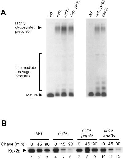
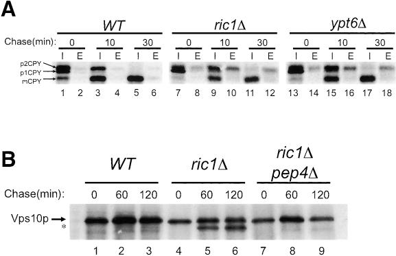
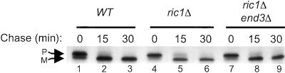
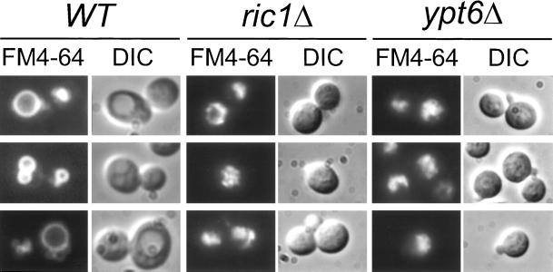
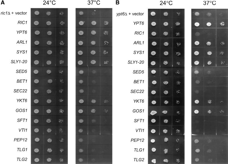
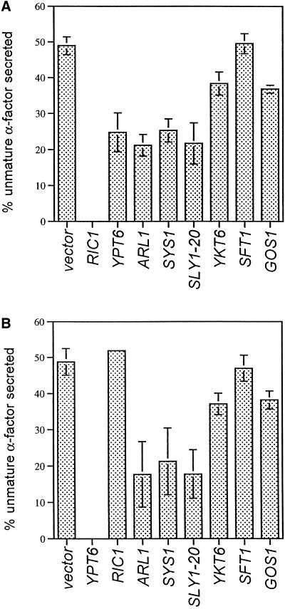
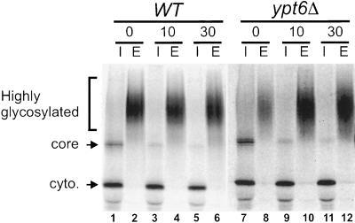
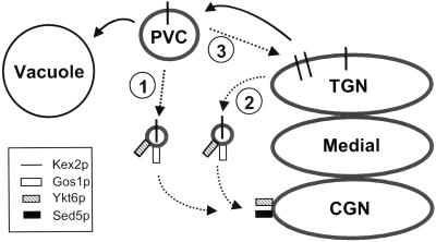
In Saccharomyces cerevisiae, clathrin is necessary for localization of trans-Golgi network (TGN) membrane proteins, a process that involves cycling of TGN proteins between the TGN and endosomes. To characterize further TGN protein localization, we applied a screen for mutations that cause severe growth defects in combination with a temperature-sensitive clathrin heavy chain. This screen yielded a mutant allele of RIC1. Cells carrying a deletion of RIC1 (ric1Delta) mislocalize TGN membrane proteins Kex2p and Vps10p to the vacuole. Delivery to the vacuole occurs in ric1Delta cells also harboring end3Delta to block endocytosis, indicative of a defect in retrieval to the TGN rather than sorting to endosomes. SYS1, originally discovered as a multicopy suppressor of defects caused by the absence of the Rab GTPase YPT6, was identified as a multicopy suppressor of ric1Delta. Further comparison of ric1Delta and ypt6Delta cells demonstrated identical phenotypes. Multicopy plasmids expressing v-SNAREs Gos1p or Ykt6p, but not other v- and t-SNAREs, partially suppressed phenotypes of ric1Delta and ypt6Delta cells. SLY1-20, a dominant activator of the cis-Golgi network t-SNARE Sed5p, also functioned as a multicopy suppressor. Because Gos1p and Ykt6p interact with Sed5p, these results raise the possibility that TGN membrane protein localization requires Ric1p- and Ypt6p-dependent retrieval to the cis-Golgi network.


Figures

Figure 1
Figure 1
Pheromone α-factor maturation and Kex2p stability are defective in ric1Δ, ypt6Δ, and gos1Δ cells. (A) Maturation of α-factor. Wild-type (WT, SEY6210), ric1Δ (GPY1480), ypt6Δ (GPY1700), ric1Δ ypt6Δ (GPY1708) and gos1Δ (GPY2108) strains were grown in SDYE media overnight at 30°C to midlogarithmic phase. Cells were labeled for 45 min at 30°C with [35S]methionine/cysteine. α-Factor was immunoprecipitated from the culture supernatant and subjected to SDS-PAGE and autoradiography. B) Kex2p stability. Wild-type (WT, SEY6210), ric1Δ (GPY1480), ric1Δ pep4Δ (GPY1608), and ric1Δ end3Δ (GPY1609) strains were grown overnight in SDYE media at 30°C to midlogarithmic phase. Cells were labeled with [35S]methionine/cysteine at 30°C for 15 min followed by the addition of excess nonlabeled amino acids (chase). Samples were removed at 0, 45, and 90 min after the addition of chase, and Kex2p was immunoprecipitated from cell lysates. Kex2p was visualized by SDS-PAGE followed by autoradiography.
Figure 2
Figure 2
CPY sorting and Vps10p stability are defective in ric1Δ and ypt6Δ strains. (A) CPY sorting. Wild-type (WT, SEY6210), ric1Δ (GPY1701) and ypt6Δ (GPY1700) strains were grown and subjected to pulse-chase analysis as described in the legend to Figure 1B. After a 15-min labeling period, samples were removed after 0, 10, and 30 min of chase and CPY was immunoprecipitated from internal (I) and external (E) fractions. CPY was resolved by SDS-PAGE and subjected to phophorimage analysis. (B) Vps10p stability. Wild-type (WT, SEY6210), ric1Δ (GPY1480), and ric1Δ pep4Δ (GPY1492) strains were grown and subjected to pulse-chase analysis as described in the legend to Figure 1B. Following a 10-min labeling period, cells were removed after 0, 60, and 120 min of chase and Vps10p was immunoprecipitated from cell lysates. Vps10p was visualized by SDS-PAGE followed by autoradiography. The 170-kDa Vps10p proteolytic cleavage product is denoted by an asterisk.
Figure 3
Figure 3
Vps10p is mislocalized in ric1Δ cells. Wild-type (WT, SEY6210) and ric1Δ (GPY1408) cells were grown at 30°C, converted to spheroplasts, and subjected to differential centrifugation. S2 and P2 are, respectively, the supernatant and pellet fractions from centrifugation at 10,000 × g for 15 min.; S3 and P3 are, respectively, the supernatant and pellet fractions from centrifugation at 200,00 × g for 17 min. Pellets were resuspended in volumes equal to the supernatants, and equal volumes were analyzed by SDS-PAGE and immunoblotting.
Figure 4
Figure 4
Efficient transport of ALP to the vacuole in ric1Δ and ric1Δ end3Δ cells. Wild-type (WT, SEY6210), ric1Δ (GPY1480), and ric1Δ end3Δ (GPY1609) strains were grown and subjected to pulse-chase analysis as described in the legend to Figure 1B. After a 10-min labeling period, cells were removed at 0, 15, and 30 min after the addition of chase, and ALP was immunoprecipitated from cell lysates. ALP was visualized by SDS-PAGE followed by autoradiography. P and M, precursor and mature forms of ALP, respectively.
Figure 5
Figure 5
ric1Δ and ypt6Δ cells display fragmented vacuoles. Wild-type (WT, SEY6210), ric1Δ (GPY1480), and ypt6Δ (GPY1700) strains were grown overnight in SDYE media, at 30°C, to midlogarithmic phase. Cells were incubated with FM4–64 for 15 min, resuspended in fresh media devoid of dye, and allowed to internalize the dye for 45 min, at 30°C, before viewing. FM4–64, fluorescence (left); DIC, differential interference contrast optics (right).
Figure 6
Figure 6
Suppression of the growth defect of ypt6Δ and ric1Δ cells. (A) GPY1480 (ric1Δ) was transformed with a vector control (pRS426), a RIC1 centromeric plasmid (p316-RIC1), and multicopy plasmids carrying the following genes: YPT6 (p426-YPT6), ARL1 (p426-ARL1), SYS1 (pric12u1A), SLY1–20 (pSLY1–20), SED5 (pSED5), BET1 (pBET1), SEC22 (pSEC22), YKT6 (pYKT6), GOS1 (p426-GOS1), SFT1 (pSFT1-URA3), VTI1 (pVTI1), PEP12 (pCB31), TLG1 (p426-TLG1), and TLG2 (pHPD1–2). Strains were grown to saturation at 24°C in SD CAA-ura and serial dilutions of the cultures were spotted onto supplemented SD plates at 24 and 37°C. (B) GPY1700 (ypt6Δ) was transformed with a vector control (pRS426), a YPT6 centromeric plasmid (YPT6), and the same plasmids as in Figure 6A, except the multicopy YPT6 plasmid was replaced with a multicopy RIC1 plasmid (p426-RIC1).
Figure 7
Figure 7
Multicopy suppression of the ric1Δ and ypt6Δ α-factor defects. ric1Δ strains (A) carrying plasmids described in the legend to Figure 6A or ypt6Δ strains carrying plasmids described in the legend to Figure 6B were grown overnight at 30°C in SD CAA-ura to midlogarithmic phase. α-Factor was metabolically labeled and immunoprecipitated as described in Figure 1A followed by phosporimage analysis using a Molecular Dynamics PhosporImager and ImageQuaNT software. Percent of unmature α-factor was calculated by dividing the amount of highly glycosylated precursor plus intermediate cleavage products by the total amount of α-factor. Values were normalized to either the RIC1-carrying strain (A) or YPT6-carrying strain (B). Error bars were calculated on the basis of three experiments except for GOS1 suppression, which was based on two experiments.
Figure 8
Figure 8
Invertase is efficiently secreted by ypt6Δ cells. Wild-type (WT, SEY6210) and ypt6Δ (GPY1700) strains expressing SUC2 from a multicopy plasmid (pRB58) were grown overnight at 30°C to midlogarithmic phase. After a 30-min incubation in low glucose media (0.1%) to induce invertase expression, cells were metabolically labeled with [35S]methionine/cysteine followed by the addition of excess nonlabeled amino acids (chase). Samples were removed after 0, 10, and 30 min of chase, and invertase was immunoprecipitated from internal (I) and external (E) fractions. Invertase was resolved by SDS-PAGE and subjected to phosphorimage analysis. Cytoplasmic (cyto), core, and highly glycosylated mature forms of invertase are shown.
Figure 9
Figure 9
Models for Ypt6p/Ric1p-mediated retrieval of TGN membrane proteins. Dotted arrows indicate possible Ypt6-dependent pathways. CGN-targeted vesicles containing Gos1p and possibly Ykt6p may originate from the prevacuolar endosome compartment (PVC; pathway 1) or from the TGN (pathway 2). Ykt6p may act as part of a Sed5p t-SNARE rather than as a vesicle-associated v-SNARE. See text for details.

References

    1. Allan BB, Balch WE. Protein sorting by directed maturation of Golgi compartments. Science. 1999;285:63–66. - PubMed
    1. Banfield DK, Lewis MJ, Pelham HR. A SNARE-like. protein required for traffic through the Golgi complex. Nature. 1995;375:806–809. - PubMed
    1. Bankaitis VA, Johnson LM, Emr SD. Isolation of yeast mutants defective in protein targeting to the vacuole. Proc Natl Acad Sci USA. 1986;83:9075–9079. - PMC - PubMed
    1. Benedetti H, Raths S, Crausaz F, Riezman H. The END3 gene encodes a protein that is required for the internalization step of endocytosis and for actin cytoskeleton organization in yeast. Mol Biol Cell. 1994;5:1023–1037. - PMC - PubMed
    1. Bensen ES, Costaguta G, Payne GS. Synthetic genetic interactions with temperature-sensitive clathrin in Saccharomyces cerevisiae: roles for synaptojanin-like Inp53p and dynamin-related Vps1p in clathrin-dependent protein sorting at the TGN. Genetics. 2000;154:83–97. - PMC - PubMed

Publication types

MeSH terms