. 2001 Jan;12(1):27-36.
doi: 10.1091/mbc.12.1.27.

Transforming growth factor-beta1 mediates epithelial to mesenchymal transdifferentiation through a RhoA-dependent mechanism

Affiliations
Free PMC article

Transforming growth factor-beta1 mediates epithelial to mesenchymal transdifferentiation through a RhoA-dependent mechanism

N A Bhowmick et al. Mol Biol Cell. 2001 Jan.
Free PMC article

Abstract

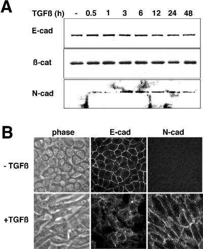
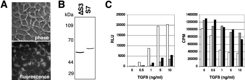
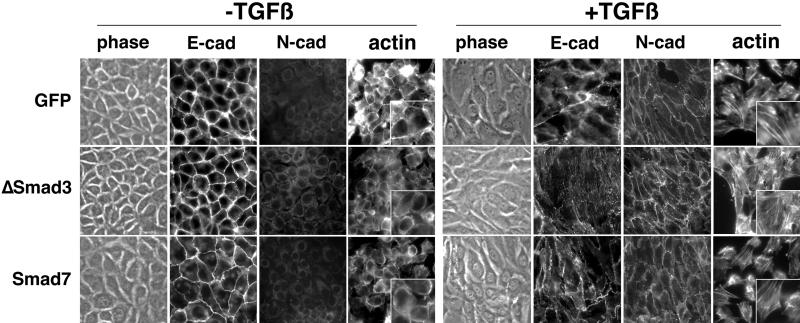
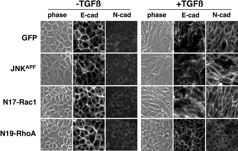
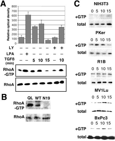
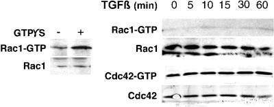
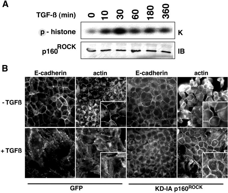
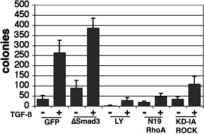
Transforming growth factor-beta1 (TGF-beta) can be tumor suppressive, but it can also enhance tumor progression by stimulating the complex process of epithelial-to-mesenchymal transdifferentiaion (EMT). The signaling pathway(s) that regulate EMT in response to TGF-beta are not well understood. We demonstrate the acquisition of a fibroblastoid morphology, increased N-cadherin expression, loss of junctional E-cadherin localization, and increased cellular motility as markers for TGF-beta-induced EMT. The expression of a dominant-negative Smad3 or the expression of Smad7 to levels that block growth inhibition and transcriptional responses to TGF-beta do not inhibit mesenchymal differentiation of mammary epithelial cells. In contrast, we show that TGF-beta rapidly activates RhoA in epithelial cells, and that blocking RhoA or its downstream target p160(ROCK), by the expression of dominant-negative mutants, inhibited TGF-beta-mediated EMT. The data suggest that TGF-beta rapidly activates RhoA-dependent signaling pathways to induce stress fiber formation and mesenchymal characteristics.


Figures

Figure 1
Figure 1
NMuMG cells treated with TGF-β undergo EMT. (A) Immunoblotting of NMuMG cells incubated with 5 ng/ml TGF-β for 0–48 h shows nearly equivalent cellular expression of E-cadherin and β-catenin through the time course, whereas N-cadherin expression is induced by 3 h of TGF-β treatment and remains elevated throughout the time course. (B) Epifluorescent and corresponding phase contrast images show that the fibroblastic cell morphology of cells treated with 5 ng/ml TGF-β for 24 h have a loss of immunodetectable E-cadherin at the cell junctions with a gain of N-cadherin expression at cell margins.
Figure 2
Figure 2
The down-regulation of SMAD signaling by retroviral transduction inhibits TGF-β–mediated responses. (A) Retroviral infection of the GFP cDNA demonstrates the high transduction efficiency of the NMuMG cells. GFP-retrovirus infected cells were observed by phase contrast and epifluorescence microscopy. (B) Expression of FLAG-tagged Smad3 or Smad7 was detected in retrovirally transduced NMuMG by immunoblotting for the epitope tag. (C) Cells stably transduced with GFP (open bar), Smad3 (gray bar), and Smad7 (closed bar) retrovirus were treated with TGF-β for 24 h and assayed for 3TP-Lux reporter activity (left) and thymidine incorporation (right).
Figure 3
Figure 3
The down-regulation of SMAD signaling does not block TGF-β–mediated EMT. NMuMG cells stably expressing GFP, Smad3, or Smad7 were treated with TGF-β (5 ng/ml for 24 h at 37°C), stained for E- or N-cadherin, and visualized by antimouse Cy2 fluorescence and phase contrast microscopy. F-actin localization was determined by Texas Red-phalloidin. The panels (400×) are magnified as insets (800×).
Figure 4
Figure 4
The expression of dominant-negative RhoA (N19-RhoA) inhibits TGF-β–mediated EMT. Forty-eight hours after retroviral infection with N19-RhoA, dominant-negative JNK (JNKAPF), dominant-negative Rac1 (N17-Rac1), or GFP cDNA as a control, NMuMG cells were treated with TGF-β (5 ng/ml for 24 h at 37°C). Phase contrast and immunofluorescent E- and N-cadherin localization was observed. Expression of dominant-negative N17-RhoA blocked TGF-β induction of EMT, whereas expression of N17-Rac1 and JNKAPF did not.
Figure 5
Figure 5
TGF-β treatment activates RhoA in epithelial cells. NMuMG cells cultured in serum-free medium were incubated with 10 ng/ml TGF-β for 0–15 min at 37°C in the presence or absence of 10 μg/ml LY294002 (LY) added 1 h before TGF-β treatment. (A) GTP loaded RhoA was measured by adsorbing cell lysates to GST-RBD beads and immunoblotted for RhoA. The densitometry measurements were normalized to total RhoA content of nonadsorbed cell lysate and represented by the bar graph (n = 4 ± SD). As a positive control, NMuMG cells treated for 5 min with LPA were assayed. TGF-β induces the rapid accumulation of RhoA-GTP in a PI3-kinase independent manner. (B) Further controls to show the GTP-loading potential of QL-RhoA– and N19-RhoA–transduced NMuMG cells assayed for RhoA activity. (C) NIH-3T3, primary keratinocytes (Pker), R1B, Mv1Lu, and BxPc3 cells were similarly assayed for RhoA activation. Only epithelial cells with intact TGF-β type I receptor exhibited TGF-β–mediated RhoA-GTP accumulation.
Figure 6
Figure 6
Rac1 and Cdc42 activation by TGF-β in NMuMG cells. TGF-β–treated (10 ng/ml for 0–60 min at 37°C) NMuMG cells were adsorbed to PBD-GST or run directly on a 12% acrylamide gel and immunoblotted sequentially for Rac1 and Cdc42. As a control, permeabilized cells incubated with GTPS were assayed for Rac1 activation (left). TGF-β–mediated induction of Rac1 and Cdc42 activation was negligible in NMuMG cells.
Figure 7
Figure 7
TGF-β regulation of p160ROCK kinase activity is involved in the EMT of NMuMG cells. (A) Myc-tagged p160ROCK was immunoprecipitated from cells treated with TGF-β (10 ng/ml for 0–360 min at 37°C) and phosphorylation of histone was resolved by electrophoreses followed by autoradiography. Expression of myc-tagged p160ROCK was determined by Western blotting. TGF-β induces p160ROCK kinase activity. (B) Cells retrovirally transduced with either GFP (as a control) or KD-IA p160ROCK cDNA were treated with 5 ng/ml TGF-β for 18 h. Subsequently, cells were fixed and stained with for E-cadherin or F-actin. The panels (400×) are magnified as insets (800×). The data suggest p160ROCK activity is involved in TGF-β–mediated actin stress fiber formation, but not E-cadherin delocalization.
Figure 8
Figure 8
TGF-β–induced motility of NMuMG cells can be antagonized by blocking the PI3-kinase or RhoA signaling pathways. Retrovirally transduced NMuMG cells allowed to attach to 8 μM polycarbonate filters (Costar, Cambridge, MA) for 18 h, were transferred to wells with fresh media supplemented with 10% serum, with or without 5 ng/ml TGF-β and/or 10 nM LY294002 (LY). The bar graph represents colonies present after 48 h in the presence or absence of 5 ng/ml TGF-β in at least two experiments done in triplicate. Blocking PI3-kinase and RhoA signaling limited TGF-β–mediated motility of NMuMG cells.

References

    1. Ashcroft GS, Yang X, Glick AB, Weinstein M, Letterio JL, Mizel DE, Anzano M, Greenwell-Wild T, Wahl SM, Deng C, Roberts AB. Mice lacking Smad3 show accelerated wound healing and an impaired local inflammatory response. Nat Cell Biol. 1999;1:260–266. - PubMed
    1. Atfi A, Djelloul S, Chastre E, Davis R, Gespach C. Evidence for a role of Rho-like GTPases and stress-activated protein kinase/c-Jun N-terminal kinase (SAPK/JNK) in transforming growth factor β-mediated signaling. J Biol Chem. 1997;272:1429–1432. - PubMed
    1. Azuma M, Tamatani T, Fukui K, Yuki T, Motegi K, Sato M. Different signaling pathways involved in transforming growth factor-β1-induced morphological change and type IV collagen synthesis in simian virus-40-immortalized normal human salivary gland duct and myoepithelial cell clones. Arch Oral Biol. 1996;41:413–424. - PubMed
    1. Benard V, Bohl BP, Bokoch GM. Characterization of rac and cdc42 activation in chemoattractant-stimulated human neutrophils using a novel assay for active GTPases. J Biol Chem. 1999;274:13198–13204. - PubMed
    1. Bishop AL, Hall A. Rho GTPases and their effector proteins. Biochem J. 2000;348:241–255. - PMC - PubMed

Publication types

MeSH terms

Substances