. 2001 Jan;12(1):53-62.
doi: 10.1091/mbc.12.1.53.

Regulation of cell cycle progression by Swe1p and Hog1p following hypertonic stress

Affiliations
Free PMC article

Regulation of cell cycle progression by Swe1p and Hog1p following hypertonic stress

M R Alexander et al. Mol Biol Cell. 2001 Jan.
Free PMC article

Abstract

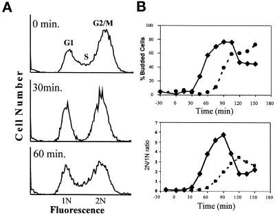
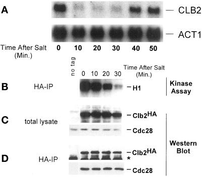
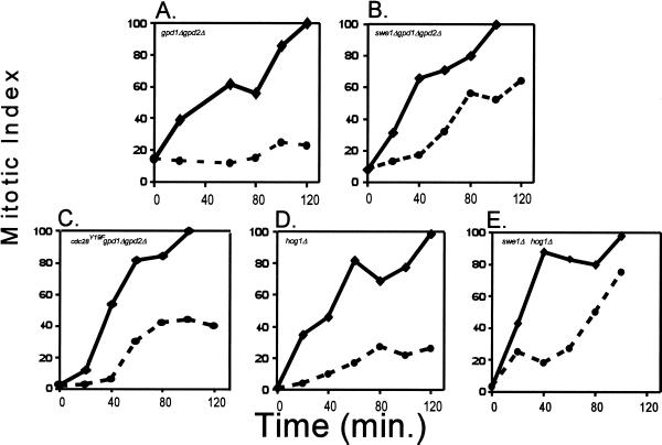
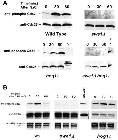
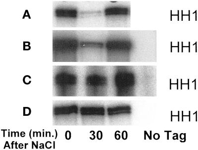
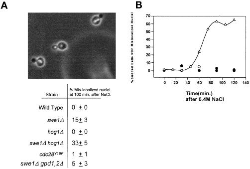
Exposure of yeast cells to an increase in external osmolarity induces a temporary growth arrest. Recovery from this stress is mediated by the accumulation of intracellular glycerol and the transcription of several stress response genes. Increased external osmolarity causes a transient accumulation of 1N and 2N cells and a concomitant depletion of S phase cells. Hypertonic stress triggers a cell cycle delay in G2 phase cells that appears distinct from the morphogenesis checkpoint, which operates in early S phase cells. Hypertonic stress causes a decrease in CLB2 mRNA, phosphorylation of Cdc28p, and inhibition of Clb2p-Cdc28p kinase activity, whereas Clb2 protein levels are unaffected. Like the morphogenesis checkpoint, the osmotic stress-induced G2 delay is dependent upon the kinase Swe1p, but is not tightly correlated with inhibition of Clb2p-Cdc28p kinase activity. Thus, deletion of SWE1 does not prevent the hypertonic stress-induced inhibition of Clb2p-Cdc28p kinase activity. Mutation of the Swe1p phosphorylation site on Cdc28p (Y19) does not fully eliminate the Swe1p-dependent cell cycle delay, suggesting that Swe1p may have functions independent of Cdc28p phosphorylation. Conversely, deletion of the mitogen-activated protein kinase HOG1 does prevent Clb2p-Cdc28p inhibition by hypertonic stress, but does not block Cdc28p phosphorylation or alleviate the cell cycle delay. However, Hog1p does contribute to proper nuclear segregation after hypertonic stress in cells that lack Swe1p. These results suggest a hypertonic stress-induced cell cycle delay in G2 phase that is mediated in a novel way by Swe1p in cooperation with Hog1p.


Figures

Figure 1
Figure 1
(A) Hypertonic stress results in a depletion of S phase cells from an asynchronous culture. Log phase cultures of wild-type MT588 were stressed by the addition of NaCl to a final concentration of 0.4 M. Samples were taken at the indicated times, and processed as described (see MATERIALS AND METHODS). The cellular DNA was stained with propidium iodide and analyzed by flow cytometry. Histograms were generated by using WinMDI software for the PC. Similar results were obtained in three separate experiments. (B) Hypertonic stress delays exit from G1. Wild-type MT588 were arrested in alpha factor, released, and stressed at time zero by the addition of 0.4 M NaCl. Budding index and ratio of 1N to 2N cells was determined for no salt control (solid line) and 0.4 M NaCl treated (dashed line) and plotted versus time in minutes after addition of NaCl.
Figure 2
Figure 2
CLB2 mRNA levels and Clb2p-Cdc28p activity decrease in response to hypertonic shock. (A) Wild-type MT588 cells were grown to log phase and stressed by the addition of NaCl to 0.4 M. Samples were taken at the indicated times. Total RNA was isolated and probed for CLB2. The same membrane was reprobed for ACT1 as a loading control. (B) Clb2p-Cdc28p kinase activity was assessed by in vitro kinase assays by using histone H1 (HH1) as a substrate. Kinase assays were performed on immunoprecipitated Clb2p-HA Cdc28p complexes isolated from wild-type MT588 cells treated the same as in A. (C) Clb2-HA and Cdc28p levels in total cell lysate used for B were determined by Western blot analysis. (D) Stability of Clb2–HA/Cdc28p complexes was determined by immunoprecipitating Clb2-HAp from the samples used for B and C. Western blots were then probed with anti-Cdc28 and anti-HA antibodies. The band marked with ∗ is IgG. W303 cells lacking the HA tag on Clb2p were used as a control. Similar results were obtained in at least three separate experiments.
Figure 3
Figure 3
Hypertonic shock causes a G2 delay. For each experiment, cells were grown and eluted in YEP + raffinose to reduce the incidence of clumping and disruption of media flow. The synchronized culture was grown until ∼80% budded. The culture was split and NaCl was added to one flask to a final concentration of 0.4 M (dashed line). The remaining flask served as a no-salt control (solid line). The mitotic index, an indication of the percentage of cells that have completed nuclear division, was determined as described (see MATERIALS AND METHODS). Time corresponds to minutes after addition of NaCl. (A) Cell cycle progression is halted when MT588 gpd1Δ gpd2Δ cells are stressed by 0.4 M NaCl. (B) swe1Δ gpd1Δ gpd2Δ cells do not halt cell cycle progression in response to osmotic stress, as indicated by the immediate increase in the mitotic index after the addition of 0.4 M NaCl. (C) Hypertonic stress causes a brief transient delay in cdc28Y19F-HA gpd1Δ gpd2Δ cells. (D) Deletion of HOG1 has a limited effect on the hypertonic stress-induced delay. (E) Response of swe1Δ hog1Δ cells to hypertonic stress was comparable to the response of swe1Δ gpd1Δ gpd2Δ cells. Similar results were obtained in at least three separate experiments.
Figure 4
Figure 4
Hypertonic stress induces the phosphorylation of Cdc28p in a Swe1p-dependent manner. Asynchronous cultures of MT588 were stressed by the addition of 0.4 M NaCl. (A) Cdc28p was precipitated with p13Suc1-agarose beads and analyzed by Western blot. Anti-phospho-Cdc2 was used to determine the phosphorylation state of Cdc28p (see MATERIALS AND METHODS). Membranes were stripped and reprobed with anti-Cdc28. A GAL: wee1+ mih1Δ strain and a Cdc28Y19F-HA strain were used as positive and negative controls, respectively. The decrease in mobility of the Y19F control is the result of the HA epitope tag. (B) Phosphorylation of Clb2-Hap-associated Cdc28p was examined by immunoprecipitating Clb2–HAp/Cdc28p complexes with anti-HA antibodies and protein A/G agarose beads. As a positive control, hyperphosphorylated Cdc28p from GAL: wee1+ mih1Δ cells was coprecipitated with p13Suc1-agarose beads. Western blots were probed with anti-phospho-Cdc2 antibodies and then stripped and reprobed with anti-Cdc28 and anti-HA antibodies. Results were consistent in three separate experiments.
Figure 5
Figure 5
Inhibition of Clb2p-Cdc28p kinase activity by exposure to 0.4 M NaCl. Wild-type MT588 (A), swe1Δ (B), hog1Δ (C), and hog1Δ swe1Δ (D) strains were grown to log phase and stressed by the addition of NaCl to 0.4 M. Clb2p–Cdc28p complexes were immunoprecipitated and assayed for activity before and after the addition of NaCl. A strain lacking the HA tag on Clb2p was used as a control. Similar results were observed in at least three independent experiments.
Figure 6
Figure 6
(A) Mislocalization of nuclei in cells that are unable to delay in G2. Cells were treated as described in Figure 3. 4,6-Diamidino-2-phenylindole was used to stain the nuclei (swe1Δ hog1Δ cells shown). The same samples were also used to determine mitotic indices (Figure 3). The percentage of cells with separated nuclei that had not properly segregated (both nuclei in the mother cell) was determined ±SD from a minimum of three independent experiments. (B) Nuclear segregation is abnormal in hog1Δ cells following salt stress after release from alpha mating factor. Wild-type and hog1Δ cultures were arrested in G1 with alpha factor and released into fresh media. After 100 min, the culture was stressed by the addition of 0.4 M NaCl. The appearance of mislocalized nuclei in budded cells following the addition of NaCl was plotted versus time. A representative experiment with wild-type cells with or without salt (○ and ●, respectively) and hog1Δ cells with and without salt (▵ and ▴, respectively) is shown here.

References

    1. Albertyn J, Hohmann S, Prior BA. Characterization of the osmotic-stress response in Saccharomyces cerevisiae: osmotic stress and glucose repression regulate glycerol-3-phosphate dehydrogenase independently. Curr Genet. 1994a;25:12–18. - PubMed
    1. Albertyn J, Hohmann S, Thevelein JM, Prior BA. GPD1, which encodes glycerol-3-phosphate dehydrogenase, is essential for growth under osmotic stress in Saccharomyces cerevisiae, and its expression is regulated by the high-osmolarity glycerol response pathway. Mol Cell Biol. 1994b;14:4135–4144. - PMC - PubMed
    1. Amon A, Tyers M, Futcher B, Nasmyth K. Mechanisms that help the yeast cell cycle clock tick: G2 cyclins transcriptionally activate G2 cyclins and repress G1 cyclins. Cell. 1993;74:993–1007. - PubMed
    1. Ansell R, Granath K, Hohmann S, Thevelein JM, Adler L. The two isoenzymes for yeast NAD+-dependent glycerol 3-phosphate dehydrogenase encoded by GPD1 and GPD2 have distinct roles in osmoadaptation and redox regulation. EMBO J. 1997;16:2179–2187. - PMC - PubMed
    1. Banuett F. Signaling in the yeasts: an informational cascade with links to the filamentous fungi. Microbiol Mol Biol Rev. 1998;62:249–274. - PMC - PubMed

Publication types

MeSH terms

Substances