Comparative Study
. 2001 Feb 1;29(3):638-43.
doi: 10.1093/nar/29.3.638.

KH domain: one motif, two folds

Affiliations
Comparative Study

KH domain: one motif, two folds

N V Grishin. Nucleic Acids Res. .

Abstract

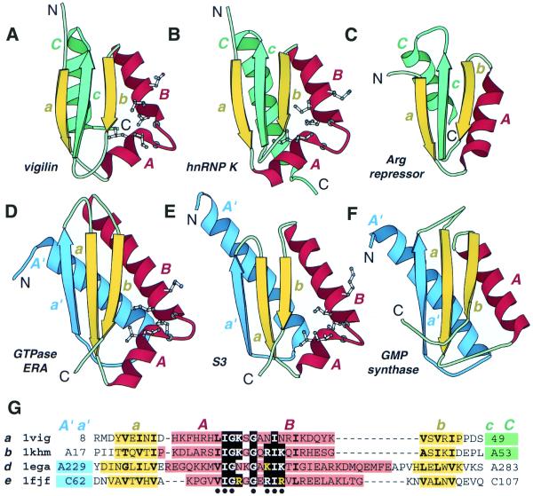
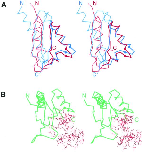
The K homology (KH) module is a widespread RNA-binding motif that has been detected by sequence similarity searches in such proteins as heterogeneous nuclear ribonucleoprotein K (hnRNP K) and ribosomal protein S3. Analysis of spatial structures of KH domains in hnRNP K and S3 reveals that they are topologically dissimilar and thus belong to different protein folds. Thus KH motif proteins provide a rare example of protein domains that share significant sequence similarity in the motif regions but possess globally distinct structures. The two distinct topologies might have arisen from an ancestral KH motif protein by N- and C-terminal extensions, or one of the existing topologies may have evolved from the other by extension, displacement and deletion. C-terminal extension (deletion) requires ss-sheet rearrangement through the insertion (removal) of a ss-strand in a manner similar to that observed in serine protease inhibitors serpins. Current analysis offers a new look on how proteins can change fold in the course of evolution.


Figures

Figure 1
Figure 1
Structural comparison of KH domains. Ribbon diagrams of type I (maxi) KH domains (A and B), type II (mini) KH domains (D and B) and non-KH proteins (C and F) were drawn by Bobscript (36), a modified version of Molscript (37). The structures were superimposed and then separated for clarity. N- and C-termini are labeled. The spatially equivalent structural elements are colored correspondingly. N- and C-terminal extension in type II (mini) and type I (maxi) KH domains and their structural equivalencies in non-KH proteins of similar fold are colored in blue and green, respectively. α-Helices and β-strands are labeled in upper and lower case italic letters, respectively. Letter color matches the color of the secondary structure element. Side chains (Cα atoms for Gly) of residues conserved in KH domains are displayed. (A) Repeat 6 of vigilin [PDB (29) entry 1VIH, residues 7–76]; (B) C-terminal KH domain of hnRNP K (PDB entry 1KHM, residues A11–A89); (C) C-terminal domain of E.coli arginine repressor (PDB entry 1XXA, residues A92–A152, the first β-strand is not shown); (D) C-terminal domain of GTPase ERA (PDB entry 1EGA, residues A186–A283); (E) N-terminal domain of ribosomal protein S3 (PDB entry 1FJF, residues C24–C106); (F) C-terminal domain of E.coli GMP synthetase (PDB entry 1GPM, residues A416–A523); (G) structure-based sequence alignment of KH motif regions from structures shown in (A, B, D and E). The panel label, PDB entry name, starting and ending residue numbers are given for each protein. Color shading and labels of secondary structure elements correspond to those in A–F. Highly conserved residues in KH domains (KH signature) are boxed with black. Hydrophobic amino acids in buried sites are shown in bold letters. Blue and green shading points to the presence of N- and C-terminal extensions whose sequences are not shown. The residues with side chains (Cα atoms for Gly) displayed on ribbon diagrams are marked with a black dot below the alignment.
Figure 2
Figure 2
Stereo diagrams of KH domains. The Cα traces of proteins are shown and the Cα atoms of the two conserved glycines in KH motif signature region are displayed as balls. N- and C-termini are labeled. (A) Stereo diagram of superimposed Cα traces of type I (maxi) KH of vigilin (red, PDB entry 1VIH, residues 6–76) and type II (mini) KH of ribosomal protein S3 (blue, PDB entry 1FJF, residues C28–C108). Superposition was performed using Insight II package (MSI). The regions used in RMSD minimization are outlined in darker colors and thicker lines. The RMSD is 2.4 Å. (B) Stereo diagram of Nova-2 KH domain (green) bound to RNA (red), PDB entry 1EC6, residues A4–A90, RNA chain C.

References

    1. Chothia C. and Lesk,A.M. (1986) The relation between the divergence of sequence and structure in proteins. EMBO J., 5, 823–826. - PMC - PubMed
    1. Hubbard T.J. and Blundell,T.L. (1987) Comparison of solvent-inaccessible cores of homologous proteins: definitions useful for protein modelling. Protein Eng., 1, 159–171. - PubMed
    1. Flores T.P., Orengo,C.A., Moss,D.S. and Thornton,J.M. (1993) Comparison of conformational characteristics in structurally similar protein pairs. Protein Sci., 2, 1811–1826. - PMC - PubMed
    1. Grishin N.V. (1997) Estimation of evolutionary distances from protein spatial structures. J. Mol. Evol., 45, 359–369. - PubMed
    1. Doolittle R.F. (1981) Similar amino acid sequences: chance or common ancestry? Science, 214, 149–159. - PubMed

Publication types

MeSH terms