. 2001 Feb;125(2):539-55.
doi: 10.1104/pp.125.2.539.

An investigation of the storage and biosynthesis of phenylpropenes in sweet basil

Affiliations

An investigation of the storage and biosynthesis of phenylpropenes in sweet basil

D R Gang et al. Plant Physiol. 2001 Feb.

Abstract

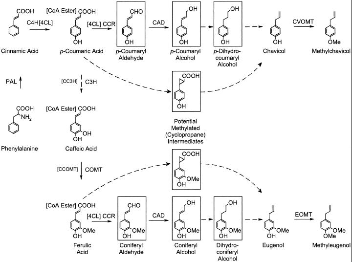
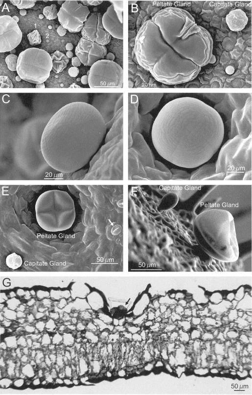
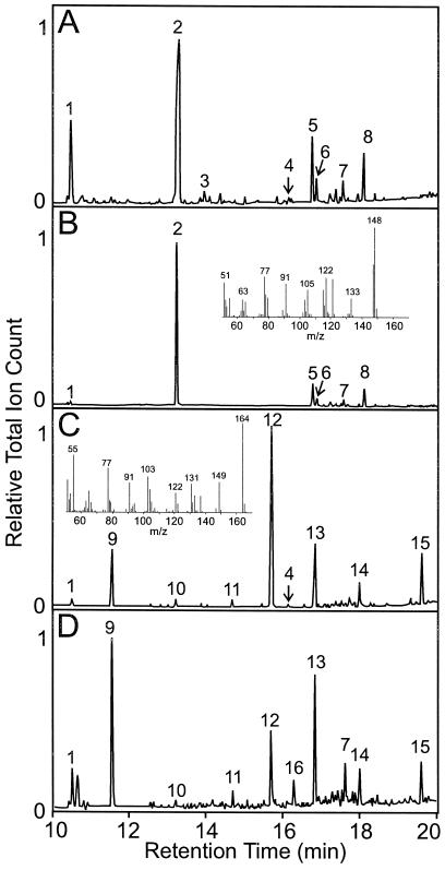
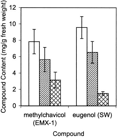
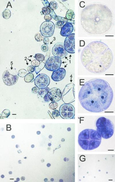
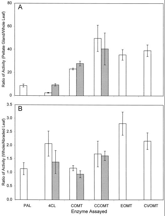
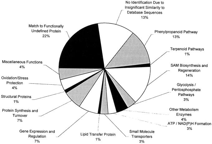
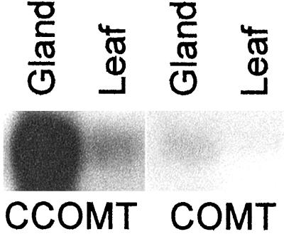
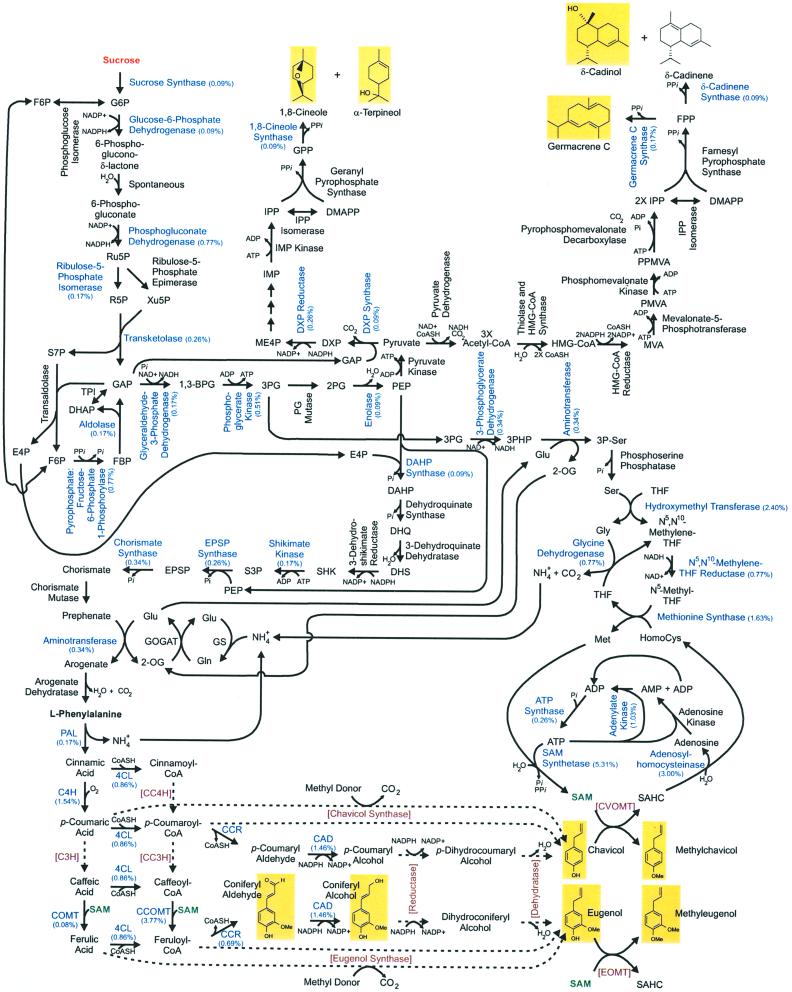
Plants that contain high concentrations of the defense compounds of the phenylpropene class (eugenol, chavicol, and their derivatives) have been recognized since antiquity as important spices for human consumption (e.g. cloves) and have high economic value. Our understanding of the biosynthetic pathway that produces these compounds in the plant, however, has remained incomplete. Several lines of basil (Ocimum basilicum) produce volatile oils that contain essentially only one or two specific phenylpropene compounds. Like other members of the Lamiaceae, basil leaves possess on their surface two types of glandular trichomes, termed peltate and capitate glands. We demonstrate here that the volatile oil constituents eugenol and methylchavicol accumulate, respectively, in the peltate glands of basil lines SW (which produces essentially only eugenol) and EMX-1 (which produces essentially only methylchavicol). Assays for putative enzymes in the biosynthetic pathway leading to these phenylpropenes localized many of the corresponding enzyme activities almost exclusively to the peltate glands in leaves actively producing volatile oil. An analysis of an expressed sequence tag database from leaf peltate glands revealed that known genes for the phenylpropanoid pathway are expressed at very high levels in these structures, accounting for 13% of the total expressed sequence tags. An additional 14% of cDNAs encoded enzymes for the biosynthesis of S-adenosyl-methionine, an important substrate in the synthesis of many phenylpropenes. Thus, the peltate glands of basil appear to be highly specialized structures for the synthesis and storage of phenylpropenes, and serve as an excellent model system to study phenylpropene biosynthesis.


Figures

Figure 1
Figure 1
Proposed biosynthetic pathway to phenylpropenes in basil. Enzymes are as follows: C4H, cinnamate 4-hydroxylase; C3H, p-coumarate 3-hydroxylase; CC3H, p-coumaroyl-CoA 3-hydroxylase; CCR, cinnamoyl-CoA reductase; and CAD, cinnamyl alcohol dehydrogenase. Dotted lines indicate hypothetical reactions; hypothetical intermediates are boxed. The phenylpropanoid pathway involving methylations of the CoA esters (as opposed to the free acids) is shown in brackets.
Figure 2
Figure 2
Morphology of basil peltate and capitate glands. A, SEM of adaxial surface of a very young EMX-1 leaf (<0.5 cm long) showing the high density at initiation of peltate and capitate glands on basil leaves. 1, Peltate gland early in development; 2, gland primordium. B, Closer view of peltate and capitate glands from same leaf as in A. The 4- and 2-fold symmetries, respectively, of each gland type are clearly visible, as are two perpendicular creases and the wrinkled folds of the immature oil sac membrane. C, Side view of a fully expanded peltate gland on a young EMX-1 leaf (1.5 cm long). D, Peltate gland on the surface of a young EMX-1 leaf, showing the fine structure of the oil sac, including the two perpendicular remnant creases left by the four underlying secretory cells. E, Adaxial surface of an expanding EMX-1 leaf (2 cm long). The peltate gland, whose oil sac is partially deflated, is recessed into the surface of the leaf. Stomatal guard cells are visible (arrow). F, Structure of glands on the surface of developing sepals from line SW, revealing the stalk cell connecting each gland to the leaf. Several non-glandular trichomes can be seen in the background. Again, the oil sac on the peltate gland is partially deflated. G, Light micrograph of an EMX-1 leaf cross-section (adaxial surface up) stained with toluidine blue showing how deeply the peltate glands (arrow) are recessed into the leaf.
Figure 3
Figure 3
GC analysis of essential oil constituents from developing leaves from basil lines EMX-1 (A) and SW (C) and from peltate glands (B and D) isolated from these two lines, respectively. Mass spectra inserted into B and C are for the major peaks, 2 and 12, respectively. Major essential oil components that were identified by in-line mass spectrometry are labeled by number: 1, cineole; 2, methylchavicol; 3, chavicol; 4, methyleugenol; 5, β-caryophyllene; 6, β-farnesene; 7, germacrene isomer I; 8, humulene; 9, linalool; 10, α-terpineol; 11, fenchyl acetate; 12, eugenol; 13, α-bergamotene; 14, germacrene isomer II; 15, δ-cadinol; and 16, β-elemene.
Figure 4
Figure 4
Effect of abrasive treatment to remove glands on content of the major phenylpropene in the essential oil of young basil leaves (1–2 cm in length) from lines EMX-1 (for methylchavicol) and SW (for eugenol). White bar, Non-treated leaves; diagonal-line filled bar, leaves rinsed with 100% (w/v) ethanol; cross-hatch filled bar, leaves from which the glands had been removed by manual abrasion and then rinsed with 100% (w/v) ethanol. Extracts from ground individual leaves, after the appropriate indicated treatment, were analyzed by GC/MS for essential oil content. Values, in milligrams of phenylpropene per gram of fresh leaf weight, are averages from leaves from five plants. Error bars are se of the mean.
Figure 5
Figure 5
Light micrographs of basil glands isolated from line EMX-1. A, Mixture of glandular trichomes prior to final purification step, showing the difference in morphology between the peltate (1) and capitate gland (2) types in basil. Some non-glandular hairs are also visible (3), as are oil sac ghosts (4) and a few broken glands (5). Scale = 20 μm. B, Purity of the peltate glands after final purification, scale = 80 μm. C and D, Isolated peltate glands, not stained with toluidine blue, with focus set at the interface between the gland disc cells and the overlying oil sac (C) and through the middle of the disc of secretory cells (D), scale = 20 μm. E and F, Isolated peltate glands stained with toluidine blue, showing the prominent nucleoli (E) and a comparison of the cross-sectional and transverse views of the disc of secretory cells (F), scale = 20 μm. G, Purity of the capitate glands after final purification, scale = 80 μm.
Figure 6
Figure 6
Peltate glandular trichomes in basil lines EMX-1 and SW are highly enriched for enzymes in the phenylpropanoid pathway. A, Comparison of the ratios of enzymatic-specific activities present in crude extracts from peltate glands with specific activities present in crude extracts from whole young leaves. B, Comparison of enzymatic-specific activities present in crude extracts from whole young leaves with specific activities present in crude extracts from young leaves with leaves abraded to remove glands. White bar, EMX-1; diagonal-line filled bar, SW.
Figure 7
Figure 7
Abundance of physiological functional classes identified in the basil peltate gland EST database.
Figure 8
Figure 8
Northern-blot analysis of the relative abundance of mRNA transcripts for CCOMT and COMT in the peltate glandular trichomes (Gland) and young leaves (Leaf) of basil line EMX-1. Ethidium bromide staining (not shown) verified that equal amounts of total RNA (4 μg) were loaded per lane in the gel.
Figure 9
Figure 9
Network of metabolic pathways leading from Suc (orange, top left) to the volatile phenylpropanoids, phenylpropenes, and terpenoids formed in EMX-1 basil peltate glands (outlined with yellow backgrounds). Blue, Enzymes for which cDNAs have been identified in the basil peltate gland EST database, with the relative proportions in the database indicated in parentheses. Black, Known enzymes for which cDNAs have not yet been identified in the database. Red with brackets, Proposed enzymes in phenylpropene pathway for which genes are yet to be identified. Dashed lines, Hypothesized conversions. Green, SAM, an important substrate for phenylpropene biosynthesis.

References

    1. Adams S, Weidenborner M. Mycelial deformations of Cladosporium herbarum due to the application of eugenol or carvacrol. J Essential Oil Res. 1996;8:535–540.
    1. Allona I, Quinn M, Shoop E, Swope K, St. Cyr S, Carlis J, Riedl J, Retzel E, Campbell MM, Sederoff R, Whetten RW. Analysis of xylem formation in pine by cDNA sequencing. Proc Natl Acad Sci USA. 1998;95:9693–9698. - PMC - PubMed
    1. Anterola AM, van Rensburg H, van Heerden PS, Davin LB, Lewis NG. Multi-site modulation of flux during monolignol formation in loblolly pine (Pinus taeda) Biochem Biophys Res Commun. 1999;261:652–657. - PubMed
    1. Bala S, Sukul N. Systemic nematocidal effect of eugenol. Nematropica. 1987;17:219–222.
    1. Bara M, Vanetti M. Antimicrobial effect of spices on the growth of Yersinia enterocolitica. J Herb Spice Med Plant. 1995;3:51–58.

Publication types

LinkOut - more resources