. 2000 Dec;6(6):1389-99.
doi: 10.1016/s1097-2765(00)00136-2.

The combined functions of proapoptotic Bcl-2 family members bak and bax are essential for normal development of multiple tissues

Affiliations

The combined functions of proapoptotic Bcl-2 family members bak and bax are essential for normal development of multiple tissues

T Lindsten et al. Mol Cell. 2000 Dec.

Abstract

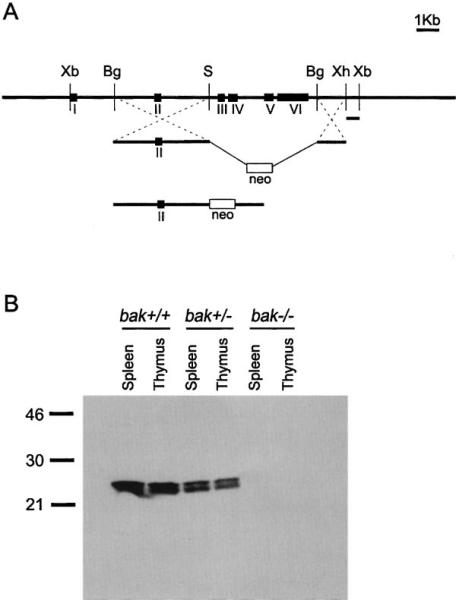
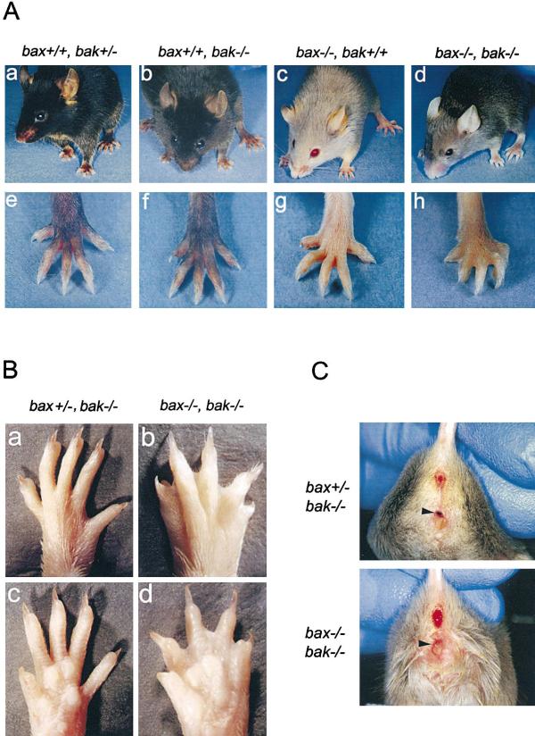
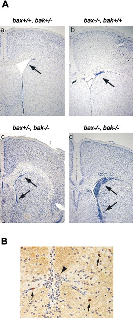
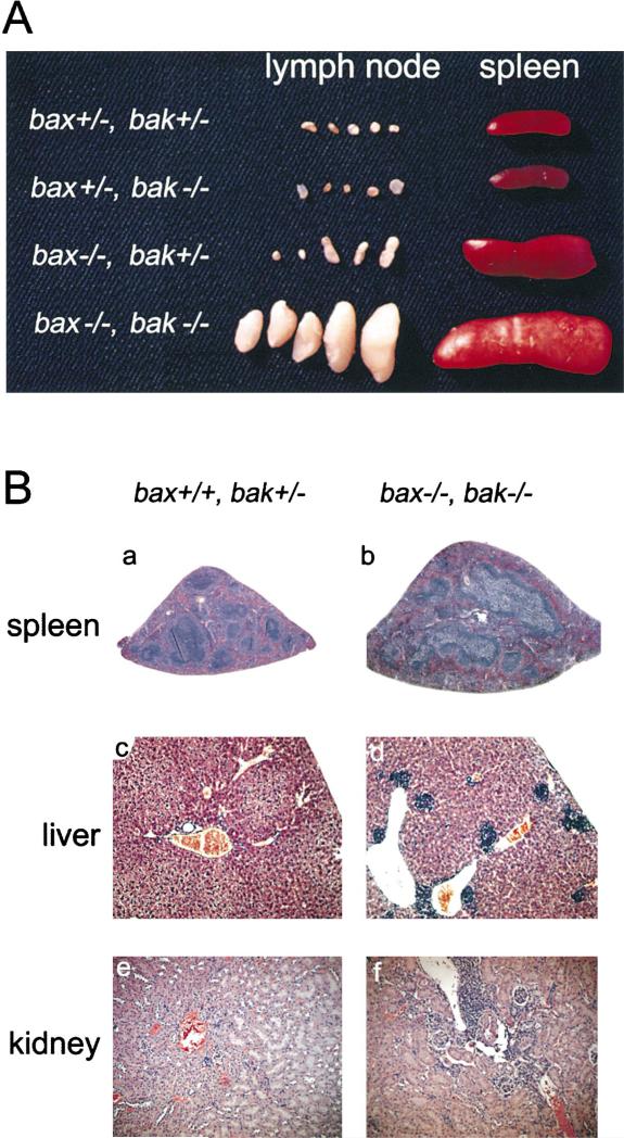
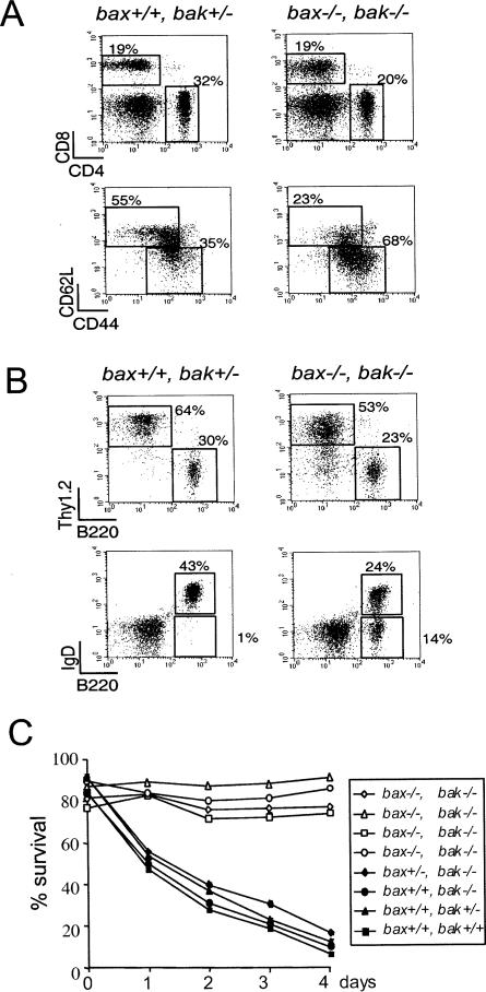
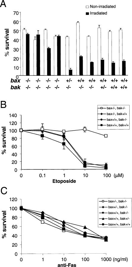
Proapoptotic Bcl-2 family members have been proposed to play a central role in regulating apoptosis. However, mice lacking bax display limited phenotypic abnormalities. As presented here, bak(-/-) mice were found to be developmentally normal and reproductively fit and failed to develop any age-related disorders. However, when Bak-deficient mice were mated to Bax-deficient mice to create mice lacking both genes, the majority of bax(-/-)bak(-/-) animals died perinatally with fewer than 10% surviving into adulthood. bax(-/-)bak(-/-) mice displayed multiple developmental defects, including persistence of interdigital webs, an imperforate vaginal canal, and accumulation of excess cells within both the central nervous and hematopoietic systems. Thus, Bax and Bak have overlapping roles in the regulation of apoptosis during mammalian development and tissue homeostasis.


Figures

Figure 1
Figure 1. Generation of bak–/– Mice
(A) The genomic organization of the bak gene, targeting strategy and the final targeting construct is shown. Restriction enzyme sites shown are (Xb), XbaI; (Bg), BglII; (S), SalI; and (Xh), XhoI. (B) Western blot analysis of whole cell lysates from spleen and thymus of bak+/+, bak+/–, and bak–/– mice.
Figure 2
Figure 2. Gross Anatomical Phenotypes of bax–/–bak–/– Mice
(A) Photographs of bax+/+bak+/– (a and e), bax+/+bak–/– (b and f), bax–/–bak+/+ (c and g), and bax–/–bak–/– (d and h) mice. (B) Dorsal and ventral views of paws from bax+/–bak–/– (a and c) and bax–/–bak–/– (b and d). (C) Photographs of vaginal openings from bax+/–bak–/– and bax–/–bak–/– mice. Arrows point to external vaginal region.
Figure 3
Figure 3. Periventricular Accumulations of Small Neuronal Cells in the Brains of bax–/–bak–/– Mice
(A) Histological sections of brain tissue from bax+/+bak+/– (a), bax–/–bak+/+ (b), bax+/–bak–/– (c), and bax–/–bak–/– (d) mice at 25× magnification. Each set is at approximately the same level and includes the anterior basal ganglia and the corpus callosum. Arrow points to the periventricular region in all sections. (B) Histological section of the dorsal midline of the mesencephalon from bax–/–bak–/– mouse at a 400× magnification, stained with anti-CD45RB. Arrows point to CD45+ cells. Arrowhead points to abnormal accumulation of small neuronal cells.
Figure 4
Figure 4. Splenomegaly, Lymphadenopathy, and Lymphoid Infiltration of Peripheral Organs in bax–/–bak–/– Mice
(A) Photograph of spleens and lymph nodes from bax–/–bak–/– and control mice. (B) Tissue sections of spleens (a and b) at 2× magnification, livers (c and d) at 10× magnification, and kidneys (e and f) at 10× magnification from bax+/+bak+/– (a, c, and e) and bax–/–bak–/– (b, d, and f) mice.
Figure 5
Figure 5. Peripheral Lymphocytes that Accumulate in bax–/–bak–/– Mice Are Enriched in Memory Cells and Fail to Die by Neglect Peripheral lymphocytes from lymph node cells were analyzed by flow cytometry
(A and B) CD4 and CD8 expression (A) and Thy1.2 and B220 expression (B). Gated T cells were also analyzed for the expression of CD44 and CD62L. Gated B cells were also analyzed for expression of IgD. (C) Peripheral lymphocytes from spleens of bax–/–bak–/– (open symbols) and control mice (closed symbols) were cultured for 4 days. Cells were stained with PI and subjected to FACS analysis at the indicated times. Samples were assayed in triplicate.
Figure 6
Figure 6. Thymocyte Death Assays in bax–/–bak–/– Mice
(A) Thymocytes from bax–/–bak–/– mice and control mice were treated with 500 rad of γ irradiation (closed bars) or left untreated (open bars). Cells were stained with PI and subjected to FACS analysis after 24 hr of culture. Mean and standard deviations of triplicate samples from each mouse are presented. (B) Thymocytes were cultured with the indicated amounts of etoposide. Cells were stained with PI and subjected to FACS analysis after 24 hr of culture. Mean and standard deviations of triplicate samples are presented. (C) Thymocytes were cultured with indicated amounts of anti-Fas antibody. Cells were stained with PI and subjected to FACS analysis after 24 hr of culture. Mean and standard deviations of triplicate samples are presented.

References

    1. Adams JM, Cory S. The Bcl-2 protein family: arbiters of cell survival. Science. 1998;281:1322–1326. - PubMed
    1. Adelman DM, Maltepe E, Simon MC. Multilineage embryonic hematopoiesis requires hypoxic ARNT activity. Genes Dev. 1999;13:2478–2483. - PMC - PubMed
    1. Alagramam KN, Kwon HY, Cacheiro NL, Stubbs L, Wright CG, Erway LC, Woychik RP. A new mouse insertional mutation that causes sensorineural deafness and vestibular defects. Genetics. 1999;152:1691–1699. - PMC - PubMed
    1. Bayer SA, Altman J. Neocortical development. Raven Press; New York: 1991.
    1. Cecconi F, Alvarez-Bolado G, Meyer BI, Roth KA, Gruss P. Apaf1 (CED-4 homolog) regulates programmed cell death in mammalian development. Cell. 1998;94:727–737. - PubMed

Publication types

MeSH terms