

Activation of frontal premotor areas during suprathreshold transcranial magnetic stimulation of the left primary sensorimotor cortex: a glucose metabolic PET study
- PMID: 11170307
- PMCID: PMC6871986
- DOI: 10.1002/1097-0193(200103)12:3<157::aid-hbm1012>3.0.co;2-v
Activation of frontal premotor areas during suprathreshold transcranial magnetic stimulation of the left primary sensorimotor cortex: a glucose metabolic PET study
Abstract
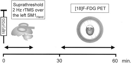
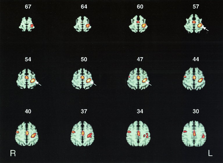
We employed cerebral (18)Fluoro-deoxyglucose positron emission tomography ([(18)F]FDG-PET) to visualize neuronal activation of the frontal motor and premotor cortex during suprathreshold repetitive transcranial magnetic stimulation (rTMS) applied to the left primary sensorimotor hand area (SM1(HAND)). Twelve right-handed normal subjects underwent two [(18)F]FDG-PET measurements at baseline without rTMS and during suprathreshold 2 Hz rTMS of the left SM1(HAND). In the rTMS condition, 1,800 magnetic stimuli at an intensity of 140% of motor-resting threshold were delivered immediately after intravenous injection of [(18)F]FDG. Relative differences in the normalized regional cerebral metabolic rate for glucose (rCMRglc) between the rTMS condition and baseline were determined using a voxel-by-voxel Student's t-test and a volume-of-interest analysis. Data analysis was a priori restricted to primary motor and premotor areas in the frontal cortex, namely the SM1, the supplementary motor area (SMA), the lateral premotor cortex (PMC), and the caudal anterior cingulate cortex (ACC) of either hemisphere. In addition to a relative increase in normalized rCMRglc in the stimulated SM1(HAND), suprathreshold rTMS was associated with well-localized increases in normalized rCMRglc in the caudal SMA and ACC on the medial wall of the frontal cortex and in the right precentral gyrus in the lateral PMC rostrally to the SM1. These data demonstrate that a selective activation of the SM1(HAND) is paralleled by an activation of a distinct set of remote premotor areas, suggesting a functional interaction between the primary motor and premotor cortex in humans.
Copyright 2001 Wiley-Liss, Inc.
Figures
Similar articles
-
Lasting cortical activation after repetitive TMS of the motor cortex: a glucose metabolic study.Neurology. 2000 Feb 22;54(4):956-63. doi: 10.1212/wnl.54.4.956. Neurology. 2000. PMID: 10690992
-
Decreased corticospinal excitability after subthreshold 1 Hz rTMS over lateral premotor cortex.Neurology. 2001 Aug 14;57(3):449-55. doi: 10.1212/wnl.57.3.449. Neurology. 2001. PMID: 11502912
-
Imaging functional activation of the auditory cortex during focal repetitive transcranial magnetic stimulation of the primary motor cortex in normal subjects.Neurosci Lett. 1999 Jul 23;270(1):37-40. doi: 10.1016/s0304-3940(99)00454-1. Neurosci Lett. 1999. PMID: 10454140
-
Left prefrontal-repetitive transcranial magnetic stimulation (rTMS) and regional cerebral glucose metabolism in normal volunteers.Psychiatry Res. 2002 Oct 1;115(3):101-13. doi: 10.1016/s0925-4927(02)00041-0. Psychiatry Res. 2002. PMID: 12208488
-
Intensity-dependent regional cerebral blood flow during 1-Hz repetitive transcranial magnetic stimulation (rTMS) in healthy volunteers studied with H215O positron emission tomography: I. Effects of primary motor cortex rTMS.Biol Psychiatry. 2003 Oct 15;54(8):818-25. doi: 10.1016/s0006-3223(03)00002-7. Biol Psychiatry. 2003. PMID: 14550681 Review.
Cited by
-
Distinct patterns of spasticity and corticospinal connectivity following complete spinal cord injury.J Physiol. 2021 Oct;599(19):4441-4454. doi: 10.1113/JP281862. Epub 2021 Sep 16. J Physiol. 2021. PMID: 34107068 Free PMC article.
-
Cortico-cortical modulation induced by 1-Hz repetitive transcranial magnetic stimulation of the temporal cortex.J Clin Neurol. 2013 Apr;9(2):75-82. doi: 10.3988/jcn.2013.9.2.75. Epub 2013 Apr 4. J Clin Neurol. 2013. PMID: 23626644 Free PMC article.
-
The supplementary motor area modulates interhemispheric interactions during movement preparation.Hum Brain Mapp. 2019 May;40(7):2125-2142. doi: 10.1002/hbm.24512. Epub 2019 Jan 17. Hum Brain Mapp. 2019. PMID: 30653778 Free PMC article.
-
Accelerated TMS - moving quickly into the future of depression treatment.Neuropsychopharmacology. 2024 Jan;49(1):128-137. doi: 10.1038/s41386-023-01599-z. Epub 2023 May 22. Neuropsychopharmacology. 2024. PMID: 37217771 Free PMC article. Review.
-
Inferring causality in brain images: a perturbation approach.Philos Trans R Soc Lond B Biol Sci. 2005 May 29;360(1457):1109-14. doi: 10.1098/rstb.2005.1652. Philos Trans R Soc Lond B Biol Sci. 2005. PMID: 16087451 Free PMC article. Review.
References
-
- Colebatch JG, Deiber MP, Passingham RE, Friston KJ, Frackowiak RSJ (1991): Regional cerebral blood flow during voluntary arm and hand movements in human subjects. J Neurophysiol 65: 1392–1401. - PubMed
-
- Cracco RQ, Amassian VE, Maccabee PJ, Cracco JB (1989): Comparison of human transcallosal resonses evoked by magnetic coil and electrical stimulation. Electroenceph Clin Neurophysiol 74: 417–424. - PubMed
-
- Dettmers C, Fink GR, Lemon RN, Stephan KM, Passingham RE, Silbersneig D, Holmes A, Ridding MC, Brooks DJ, Frackowiak RS (1995): Relation between cerebral activity and force in motor areas of human brain. J Neurophysiol 74: 802–815. - PubMed
Publication types
MeSH terms
Substances
LinkOut - more resources
Full Text Sources
