

Nerve growth factor is expressed by postmitotic avian retinal horizontal cells and supports their survival during development in an autocrine mode of action
- PMID: 11171331
- PMCID: PMC2710126
- DOI: 10.1242/dev.128.4.471
Nerve growth factor is expressed by postmitotic avian retinal horizontal cells and supports their survival during development in an autocrine mode of action
Abstract
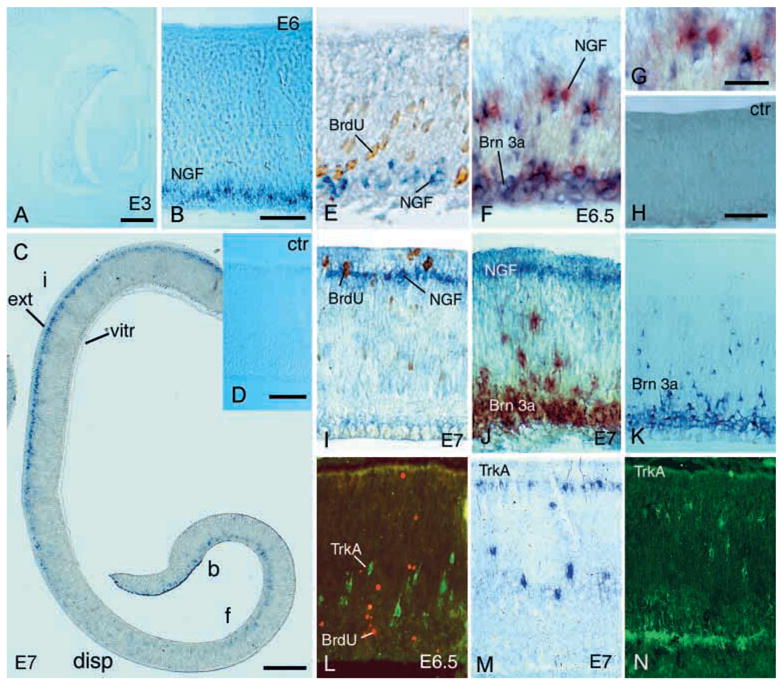
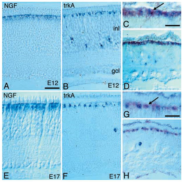
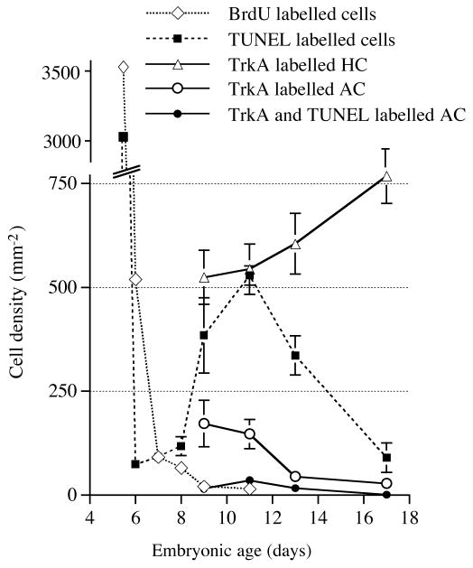
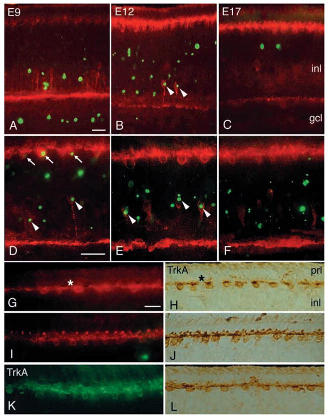
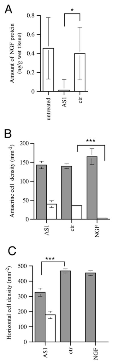
Cell death in the developing retina is regulated, but so far little is known about what factors regulate the cell death. Several neurotrophic factors and receptors, including the neurotrophins and Trk receptors, are expressed during the critical time. We have studied the developing avian retina with respect to the role of nerve growth factor (NGF) in these processes. Our starting point for the work was that NGF and its receptor TrkA are expressed in a partially overlapping pattern in the inner nuclear layer of the developing retina. Our results show that TrkA and NGF-expressing cells are postmitotic. The first NGF-expressing cells were found on the vitreal side of the central region of E5.5-E6 retina. This pattern changed and NGF-expressing cells identified as horizontal cells were later confined to the external inner nuclear layer. We show that these horizontal cells co-express TrkA and NGF, unlike a subpopulation of amacrine cells that only expresses TrkA. In contrast to the horizontal cells, which survive, the majority of the TrkA-expressing amacrine cells die during a period of cell death in the inner nuclear layer. Intraocular injections of NGF protein rescued the dying amacrine cells and injection of antisense oligonucleotides for NGF that block its synthesis, caused death among the TrkA-expressing horizontal cells, which normally would survive. Our results suggest that NGF supports the survival of TrkA expressing avian horizontal cells in an autocrine mode of action in the retina of E10-E12 chicks. The cells co-express TrkA and NGF and the role for NGF is to maintain the TrkA-expressing horizontal cells. The TrkA-expressing amacrine cells are not supported by NGF and subsequently die. In addition to the effect on survival, our results suggest that NGF plays a role in horizontal cell plasticity.
Figures
Similar articles
-
Nerve growth factor receptor TrkA is expressed by horizontal and amacrine cells during chicken retinal development.J Comp Neurol. 1998 Oct 26;400(3):408-16. J Comp Neurol. 1998. PMID: 9779944 Free PMC article.
-
Effect of p75NTR on the regulation of naturally occurring cell death and retinal ganglion cell number in the mouse eye.Dev Biol. 2006 Feb 1;290(1):57-65. doi: 10.1016/j.ydbio.2005.08.051. Epub 2005 Dec 15. Dev Biol. 2006. PMID: 16343477
-
Expression of neurotrophins and trk receptors in the avian retina.J Comp Neurol. 1996 Jan 22;364(4):664-76. doi: 10.1002/(SICI)1096-9861(19960122)364:4<664::AID-CNE5>3.0.CO;2-1. J Comp Neurol. 1996. PMID: 8821453
-
Biogenesis and function of the NGF/TrkA signaling endosome.Int Rev Cell Mol Biol. 2015;314:239-57. doi: 10.1016/bs.ircmb.2014.10.002. Epub 2014 Nov 18. Int Rev Cell Mol Biol. 2015. PMID: 25619719 Free PMC article. Review.
-
Neurotrophins in cultured cells from periodontal tissues.J Periodontol. 2003 Jan;74(1):76-84. doi: 10.1902/jop.2003.74.1.76. J Periodontol. 2003. PMID: 12593600 Review.
Cited by
-
Horizontal Cells, the Odd Ones Out in the Retina, Give Insights into Development and Disease.Front Neuroanat. 2016 Jul 19;10:77. doi: 10.3389/fnana.2016.00077. eCollection 2016. Front Neuroanat. 2016. PMID: 27486389 Free PMC article. Review.
-
The heterogenic final cell cycle of chicken retinal Lim1 horizontal cells is not regulated by the DNA damage response pathway.Cell Cycle. 2014;13(3):408-17. doi: 10.4161/cc.27200. Epub 2013 Nov 18. Cell Cycle. 2014. PMID: 24247150 Free PMC article.
-
Alternative splicing of neuroligin and its protein distribution in the outer plexiform layer of the chicken retina.J Comp Neurol. 2010 Dec 15;518(24):4938-62. doi: 10.1002/cne.22499. J Comp Neurol. 2010. PMID: 21031560 Free PMC article.
-
Characterization of NGF, trkA (NGFR) , and p75 (NTR) in Retina of Mice Lacking Reelin Glycoprotein.Int J Cell Biol. 2014;2014:725928. doi: 10.1155/2014/725928. Epub 2014 Jan 30. Int J Cell Biol. 2014. PMID: 24627687 Free PMC article.
-
Mesenchymal stem cells for retinal diseases.Int J Ophthalmol. 2011;4(4):413-21. doi: 10.3980/j.issn.2222-3959.2011.04.19. Epub 2011 Aug 18. Int J Ophthalmol. 2011. PMID: 22553693 Free PMC article.
References
-
- Acheson A, Conover JC, Fandl JP, DeChiara TM, Russell M, Thadani A, Squinto SP, Yancopoulos GD, Lindsay RM. A BDNF autocrine loop in adult sensory neurons prevents cell death. Nature. 1995;374:450–453. - PubMed
-
- Bäckström A, Söderström S, Kylberg A, Ebendal T. Molecular cloning of the chicken trkA and its expression in early peripheral ganglia. J Neurosci Res. 1996;46:67–81. - PubMed
-
- Catsicas M, Osen-Sand A, Staple J, Jones KA, Ayala G, Knowles J, Grenningloh G, Merlio-Pich E, Catsicas S. Antisense blockade of expression, SNAP-25 in vitro and in vivo. In: Agrawal S, editor. Methods in Molecular Medicine: Antisense Therapeutics. Totowa, NJ: Humana Press; 1995. pp. 57–85. - PubMed
-
- Cepko CL. The roles of intrinsic and extrinsic cues and bHLH genes in the determination of retinal cell fates. Curr Opin Neurobiol. 1999;9:37–46. - PubMed
Publication types
MeSH terms
Substances
Grants and funding
LinkOut - more resources
Full Text Sources
