. 2001 Feb 13;98(4):1513-8.
doi: 10.1073/pnas.98.4.1513. Epub 2001 Feb 6.

The surface coat of procyclic Trypanosoma brucei: programmed expression and proteolytic cleavage of procyclin in the tsetse fly

Affiliations

The surface coat of procyclic Trypanosoma brucei: programmed expression and proteolytic cleavage of procyclin in the tsetse fly

A Acosta-Serrano et al. Proc Natl Acad Sci U S A. .

Abstract

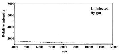
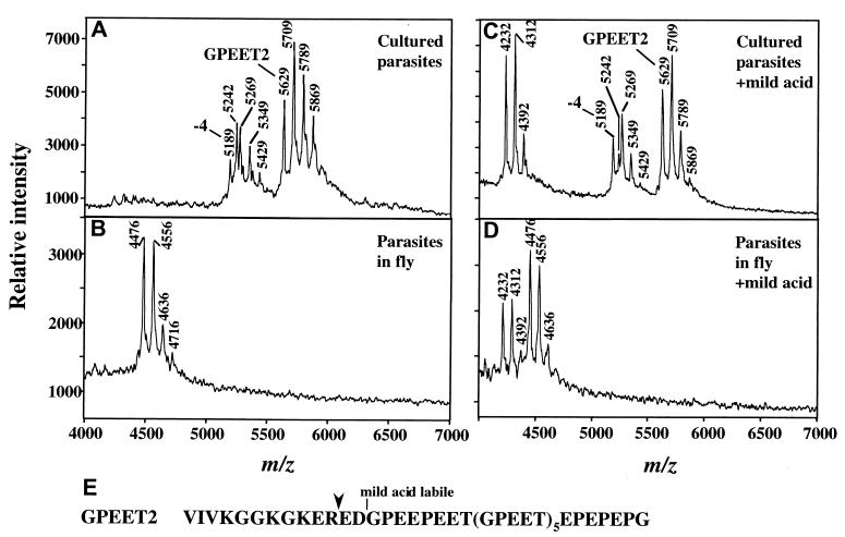
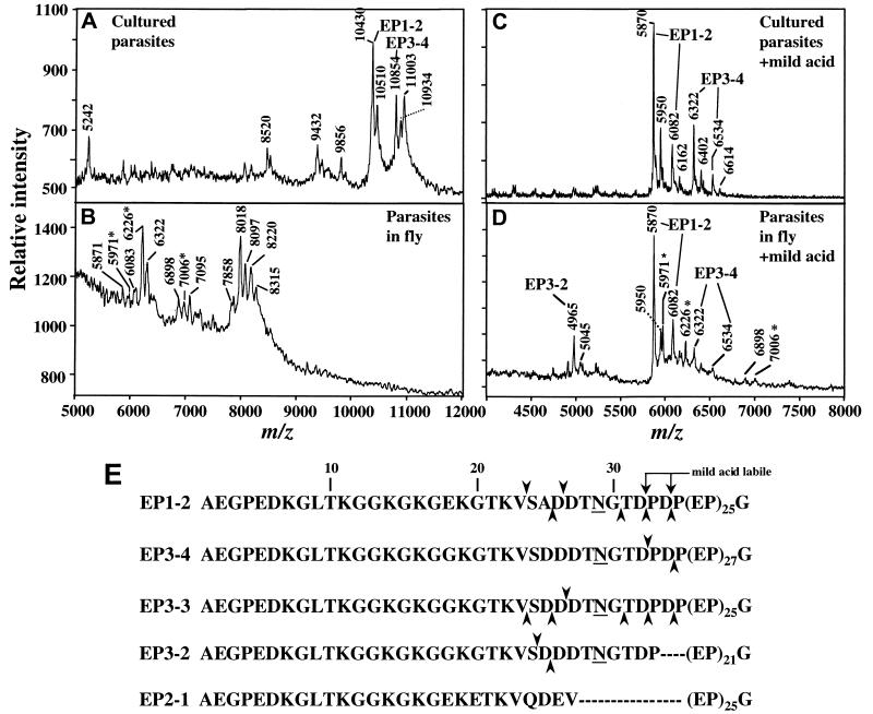
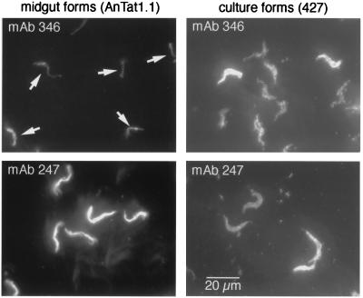
Trypanosoma brucei, the protozoan parasite causing sleeping sickness, is transmitted by a tsetse fly vector. When the tsetse takes a blood meal from an infected human, it ingests bloodstream form trypanosomes that quickly differentiate into procyclic forms within the fly's midgut. During this process, the parasite loses the 10(7) molecules of variant surface glycoprotein that formed its surface coat, and it develops a new coat composed of several million procyclin molecules. Procyclins, the products of a small multigene family, are glycosyl phosphatidylinositol-anchored proteins containing characteristic amino acid repeats at the C terminus [either EP (EP procyclin, a form of procyclin rich in Glu-Pro repeats) or GPEET (GPEET procyclin, a form of procyclin rich in Glu-Pro-Glu-Glu-Thr repeats)]. We have used a sensitive and accurate mass spectrometry method to analyze the appearance of different procyclins during the establishment of midgut infections in tsetse flies. We found that different procyclin gene products are expressed in an orderly manner. Early in the infection (day 3), GPEET2 is the only procyclin detected. By day 7, however, GPEET2 disappears and is replaced by several isoforms of glycosylated EP, but not the unglycosylated isoform EP2. Unexpectedly, we discovered that the N-terminal domains of all procyclins are quantitatively removed by proteolysis in the fly, but not in culture. These findings suggest that one function of the protease-resistant C-terminal domain, containing the amino acid repeats, is to protect the parasite surface from digestive enzymes in the tsetse fly gut.


Figures

Figure 1
Figure 1
MS of contents of uninfected fly guts. Guts were isolated 3 days after feeding with noninfected blood, and contents were treated by the procyclin extraction/GPI removal protocol. A 1-butanol extract equivalent to five guts was analyzed.
Figure 2
Figure 2
MS of procyclins from trypanosomes cultured in vitro (A and C) or from tsetse flies three days after infection (B and D). (A) Analysis of aq.HF-treated-procyclins from ≈106 parasites cultured in vitro in the presence of 10 mM glycerol. The ion at m/z 5,629 is full length GPEET2, and it is accompanied by phosphorylated species (m/z 5,709, 5,789, and 5,869). The ion at m/z 5,189 is GPEET2 procyclin truncated by 4 residues, and it is also accompanied by phosphorylated species (m/z 5,269, 5,349, and 5,429). The ion at m/z 5,242 (also present in C) is an unknown polypeptide. Low levels of EP procyclin (EP1–2 and EP3–4) are detected in the 9,000–11,000 m/z range (not shown). (B) Analysis of aq.HF treated-procyclins from ≈7 × 10⁵ parasites extracted from tsetse flies 3 days after infection. The ion at m/z 4,476 is a proteolytic fragment of GPEET2 procyclin (see E for cleavage site), and it is accompanied by ions of phosphorylated species (m/z 4,556, 4636, and 4,716). (C) Mild acid hydrolysis of aq.HF-treated procyclins from cultured trypanosomes, identical to those in A. (D) Mild acid hydrolysis of the aq.HF treated-procyclins from tsetse-derived trypanosomes, identical to those in B. Note in both C and D, the ion of m/z 4,232, representing the C-terminal fragment generated by partial mild acid cleavage at D13 (16). This ion is accompanied by phosphorylated species (m/z 4,312 and 4,392). No fragments of EP procyclins were detected in D. (E) Sequence of GPEET2 procyclin. Downward arrowhead, site of proteolytic cleavage in the fly midgut.
Figure 3
Figure 3
MS of procyclins from trypanosomes cultured in vitro (A and C) or from tsetse flies 7 days after infection (B and D). (A) Analysis of aq.HF-treated-procyclins from ≈106 parasites cultured in vitro in the absence of glycerol to promote expression of EP procyclin. Ions at m/z 10,430 and 10,854 are from full-length EP1–2 and EP3–4. Ions at m/z 10,510 and 10,934 are from the same procyclin species, except there is a residual phosphate group, derived from the GPI anchor, linked to the C-terminal ethanolamine. These phosphorylated species are not detected after longer HF treatment (11). Ions at m/z 9,432 and 9,856 are from EP1–2 and EP3–4 procyclins, respectively, which are missing 10 amino acids from the N terminus (11). The m/z ions 5,242 and 8,520 are polypeptide contaminants, and that at m/z 11,003 is probably KMP-11, a protein that frequently contaminates procyclin (33). (B) Analysis of aq.HF-treated procyclins from ≈3 × 10⁵ parasites extracted from infected tsetse flies at 7 days after infection. See Table 1 for assignments of ions. No ions derived from GPEET2 were detected. Ions with asterisks in B and D are probably contaminants (see text). (C) Mild acid hydrolysis of aq.HF-treated-procyclins, from cultured trypanosomes, identical to those in A. (D) Mild acid hydrolysis of the aq.HF-treated-procyclins, from tsetse-derived-trypanosomes, identical to those in B. Each of the C-terminal fragments after mild acid hydrolysis (C and D) is paired with a corresponding species phosphorylated on ethanolamine (with m/z increased by 80 Da). (E) Primary sequence of mature EP procyclins, with numbering starting at the N terminus of the mature protein. Upward arrowheads indicate cleavages detected at day 7. Downward arrowheads indicate cleavages detected at days 14–28 (Table 1). Underlines indicate glycosylation sites (N29).
Figure 4
Figure 4
Immunofluorescence analysis of parasites isolated from tsetse flies. Trypanosomes from tsetse flies 14 days after infection were processed for immunofluorescence and stained with antibodies specific for the EP N terminus (mAb 346) or the EP repeat sequence (mAb 247). Procyclic culture forms (427) were used as a positive control. The same exposure times and settings were used for all four images. Arrows indicate midgut-derived trypanosomes that were weakly staining for mAb 346 [the location of cells was confirmed by 4′,6-diamidino-2-phenylindole staining (not shown)]. Stock 427, which expresses high levels of GPEET (12), commonly shows heterogeneous staining with anti-EP antibodies (unpublished observations).

References

    1. Vickerman K. Br Med Bull. 1985;41:105–114. - PubMed
    1. Van Den Abbeele J, Claes Y, van Bockstaele D, Le Ray D, Coosemans M. Parasitology. 1999;118:469–478. - PubMed
    1. Ziegelbauer K, Quinten M, Schwarz H, Pearson T W, Overath P. Eur J Biochem. 1990;192:373–378. - PubMed
    1. Matthews K R, Gull K. J Cell Biol. 1994;125:1147–1156. - PMC - PubMed
    1. Roditi I, Carrington M, Turner M. Nature (London) 1987;325:272–274. - PubMed

Publication types

MeSH terms

Substances

LinkOut - more resources