. 2001 Feb 13;98(4):1589-94.
doi: 10.1073/pnas.98.4.1589.

Embryonic expression of the tumor-associated PAX3-FKHR fusion protein interferes with the developmental functions of Pax3

Affiliations

Embryonic expression of the tumor-associated PAX3-FKHR fusion protein interferes with the developmental functions of Pax3

M J Anderson et al. Proc Natl Acad Sci U S A. .

Abstract

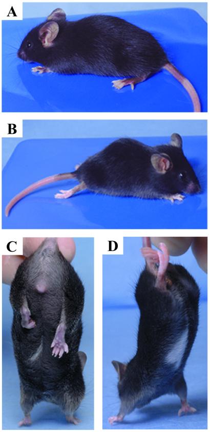
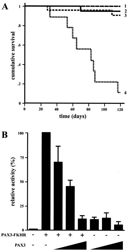
A unique chromosomal translocation involving the genes PAX3 and FKHR is characteristic of most human alveolar rhabdomyosarcomas. The resultant chimeric protein fuses the PAX3 DNA-binding domains to the transactivation domain of FKHR, suggesting that PAX3-FKHR exerts its role in alveolar rhabdomyosarcomas through dysregulation of PAX3-specific target genes. Here, we have produced transgenic mice in which PAX3-FKHR expression was driven by mouse Pax3 promoter/enhancer sequences. Five independent lines expressed PAX3-FKHR in the dorsal neural tube and lateral dermomyotome. Each line exhibited phenotypes that correlated with PAX3-FKHR expression levels and predominantly involved pigmentary disturbances of the abdomen, hindpaws, and tail, with additional neurological related alterations. Phenotypic severity could be increased by reducing Pax3 levels through matings with Pax3-defective Splotch mice, and interference between PAX3 and PAX3-FKHR was apparent in transcription reporter assays. These data suggest that the tumor-associated PAX3-FKHR fusion protein interferes with normal Pax3 developmental functions as a prelude to transformation.


Figures

Figure 1
Figure 1
Creation of PAX3-FKHR transgenic mice. (A) The two transgene constructs contain mouse Pax3 promoter/enhancer sequences upstream of the PAX3-FKHR cDNA. (B) Transgene copy number assessment by Southern blot analysis. Probe A detects an endogenous Pax3-specific band, as well as band(s) specific to the transgene. The profiles of pMP3-D13(HA) derived lines differ from MP3–5 because of a BamHI restriction site in the HA tag. Comparison between the relative band intensities allowed for copy number determination.
Figure 2
Figure 2
PAX3-FKHR transgene expression correlates with neural tube dysmorphology. (A) Whole-mount in situ hybridization shows transgene expression in the dorsal neural tube and lateral dermomyotome. Expression of mouse Pax3 in an entire control embryo is shown for comparison. With the exception of MP3–5, expression in the developing head region was absent in the other lines. (B) H&E-stained sections of E11.5 neural tubes taken at the level of the hindlimb buds.
Figure 3
Figure 3
Skeletal muscle morphology and histochemical staining in transgenic mice compared with control. (AE) H&E-stained vastus lateralis muscle. (C, D, and E) Marked variation in fiber size, increased numbers of fibers with central nuclei (arrowheads), and basophilic staining regions (arrow) were observed in a severely affected MP3–5 mouse. Staining in control (A) and each of the other lines (B, represented here with an HA21 mouse) was normal. (FH) Fiber-type grouping is shown by the acid-stable myosin ATPase reaction. In control muscle (F), type 1 (brown) fibers are uniformly distributed among type 2 (pink) fibers. This uniform staining was seen in HA21, HA39, and HA83 lines (G, represented here with an HA83h mouse). (H) Fiber-type grouping (clustering of brown-staining type 1 fibers) was found in a severely affected MP3–5 mouse. C was taken at a higher magnification than the others. (A, B, E–H, ×190; C, ×760; D, ×390.)
Figure 4
Figure 4
Analysis of neural crest cell emigration from the dorsal neural tube. Whole-mount E10.5 Wnt-1/LacZ (control) and Tg/Wnt-1/LacZ embryos stained for β-galactosidase activity. Staining of cells in the cranial region, including the branchial arches and nasotemporal region around the eyes, was similar among each of the embryos. In the posterior region, centering on the hindlimb buds, fewer labeled cells in the transgenic lines can be seen emigrating from the neural tube (arrowheads). (Lower) A higher magnification of the hindlimb bud region. For orientation, the forelimb (F) and hindlimb (H) buds are labeled. (Upper, ×7; Lower, ×15.)
Figure 5
Figure 5
Representative phenotypes. (A and C) HA21 mouse showing overall appearance. Pigmentary disturbances of the tail and hindpaws were the most common features seen in all lines, except HA83l (A). A white belly patch was observed at a relatively low frequency only in HA21 and HA39 mice (C). HA21 and affected MP3–5 mice behaved abnormally when held by their tail, by pulling their hindlimb(s) to their body (C). (B and D) Compound HA21/Splotch heterozygotes displayed severe spinal dysraphism and kyphosis, paralysis and reduced sensory perception of their hindlimbs and tail, and exaggerated gait involving dragging their hindlimbs.
Figure 6
Figure 6
Negative influence of PAX3-FKHR on Pax3 activity. Kaplan–Meier survival plots of offspring resulting from HA21 × Splotch matings show that HA21 Tg+Sp± mice had a significantly reduced lifespan (A). The survival of TgSp+/+ (row 2, n = 20), TgSp± (row 1, n = 20), Tg+Sp+/+ (row 3, n = 23), and Tg+Sp± (row 4, n = 9) mice was monitored over the course of 120 days. PAX3 and PAX3-FKHR compete for transcriptional activity (B). When increasing amounts of PAX3 were added (50 ng, 100 ng, and 1 μg), PAX3-FKHR transactivation of PRS-9 was blocked to the levels of PAX3 alone. The activity from PAX3-FKHR alone was arbitrarily set at 100%.

References

    1. Kagan J, Croce C M. Ann Oncol. 1991;2:9–21. - PubMed
    1. Rabbitts T H. Nature (London) 1994;372:143–149. - PubMed
    1. Turc-Carel C, Lizard-Nacol S, Justrabo E, Favrot M, Philip T, Tabone E. Cancer Genet Cytogenet. 1986;19:361–362. - PubMed
    1. Douglass E C, Valentine M, Etcubanas E, Parham D, Webber B L, Houghton P J, Green A A. Cytogenet Cell Genet. 1987;45:148–155. - PubMed
    1. Galili N, Davis R J, Fredericks W J, Mukhopadhyay S, Rauscher F J, III, Emanuel B S, Rovera G, Barr F G. Nat Genet. 1993;5:230–235. - PubMed

Publication types

MeSH terms