. 2001 Feb 13;98(4):1607-12.
doi: 10.1073/pnas.98.4.1607.

Dual modulation of cell survival and cell death by beta(2)-adrenergic signaling in adult mouse cardiac myocytes

Affiliations

Dual modulation of cell survival and cell death by beta(2)-adrenergic signaling in adult mouse cardiac myocytes

W Z Zhu et al. Proc Natl Acad Sci U S A. .

Abstract

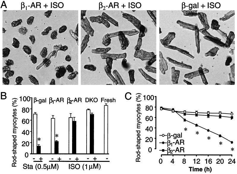
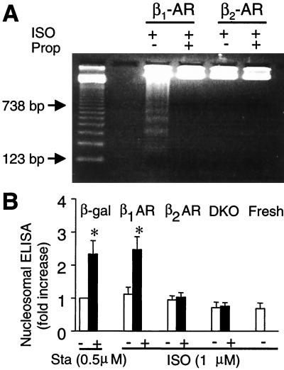
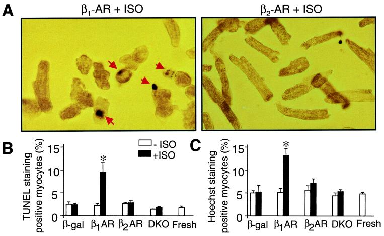
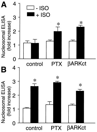
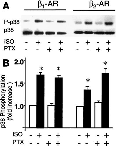
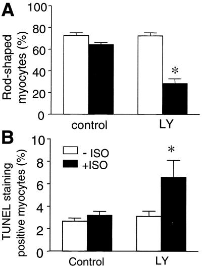
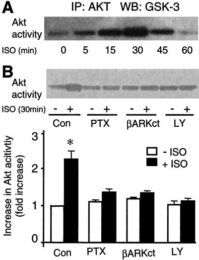
The goal of this study was to determine whether beta(1)-adrenergic receptor (AR) and beta(2)-AR differ in regulating cardiomyocyte survival and apoptosis and, if so, to explore underlying mechanisms. One potential mechanism is that cardiac beta(2)-AR can activate both G(s) and G(i) proteins, whereas cardiac beta(1)-AR couples only to G(s). To avoid complicated crosstalk between beta-AR subtypes, we expressed beta(1)-AR or beta(2)-AR individually in adult beta(1)/beta(2)-AR double knockout mouse cardiac myocytes by using adenoviral gene transfer. Stimulation of beta(1)-AR, but not beta(2)-AR, markedly induced myocyte apoptosis, as indicated by increased terminal deoxynucleotidyltransferase-mediated UTP end labeling or Hoechst staining positive cells and DNA fragmentation. In contrast, beta(2)-AR (but not beta(1)-AR) stimulation elevated the activity of Akt, a powerful survival signal; this effect was fully abolished by inhibiting G(i), G(beta gamma), or phosphoinositide 3 kinase (PI3K) with pertussis toxin, beta ARK-ct (a peptide inhibitor of G(beta gamma)), or LY294002, respectively. This indicates that beta(2)-AR activates Akt via a G(i)-G(beta gamma)-PI3K pathway. More importantly, inhibition of the G(i)-G(beta gamma)-PI3K-Akt pathway converts beta(2)-AR signaling from survival to apoptotic. Thus, stimulation of a single class of receptors, beta(2)-ARs, elicits concurrent apoptotic and survival signals in cardiac myocytes. The survival effect appears to predominate and is mediated by the G(i)-G(beta gamma)-PI3K-Akt signaling pathway.


Figures

Figure 1
Figure 1
β1-AR but not β2-AR stimulation induces loss of rod-shaped myocytes. (A) Cardiac myocytes infected by adeno-β1-AR, adeno-β2-AR, or adeno-β-gal (moi 100) were cultured for 24 h in the presence of ISO (1 μM). (B) The average percentage of rod-shaped myocytes was markedly reduced by stimulation of β1-AR but not β2-AR. Sta was used as a positive control. DKO refers to uninfected DKO cells, whereas Fresh refers to freshly isolated cells. (C) The time course of ISO-induced drop of rod-shaped cells in myocytes infected by adeno-β1-AR, adeno-β2-AR, or adeno-β-gal. *, P < 0.01 versus untreated (ISO or Sta) or uninfected myocytes (B) or those infected by adeno-β2-AR or adeno-β-gal (B and C) (n = 4–6 for each data point).
Figure 2
Figure 2
Stimulation of β1-AR but not β2-AR increases DNA fragmentation, assayed by DNA laddering (A) or nucleosomal ELISA (B). *, P < 0.01 versus untreated (Sta or ISO) or uninfected myocytes (DKO and Fresh) and those infected by adeno-β2-AR (n = 4–6 for each group).
Figure 3
Figure 3
Stimulation of β1-AR but not β2-AR induces cell apoptosis. (A) Representative micrographs show TUNEL staining in myocytes infected by adeno-β1-AR or adeno-β2-AR and treated with ISO. Apoptotic nuclei appear blue by TUNEL staining with a varying degree of condensation of nuclear chromatin (arrows). (B and C) The average data of TUNEL and Hoechst staining. *, P < 0.01 versus untreated or uninfected myocytes and those infected by adeno-β2-AR (n = 5 to 6 for each group).
Figure 4
Figure 4
Involvement of Gi and Gβγ in β2-AR-mediated survival effect. PTX treatment or coexpression of βARK-ct permits β2-AR stimulation by ISO to induce DNA fragmentation (A). These interventions neither blunt nor enhance the effect of β1-AR stimulation (B). *, P < 0.01 versus ISO-untreated myocytes (n = 4–6 for each group).
Figure 5
Figure 5
p38 MAPK is not involved in β2-AR-mediated survival effect. (A) Representative Western blots show that stimulation of β1-AR or β2-AR by ISO increases p38 MAPK phosphorylation in a PTX-insensitive manner. (B) The average data (n = 5 to 6, P < 0.01 versus ISO).
Figure 6
Figure 6
Inhibition of PI3K by LY 294002 abolishes β2-AR-induced survival effect and unmasks β2-AR-mediated apoptotic effect. β2-AR stimulation by ISO (1 μM) reduces the percentage of the rod-shaped myocytes (A) and increases the TUNEL staining positive cells (B) only in the presence of LY294002 (10 μM). *, P < 0.01 versus the groups in the absence of ISO and the group with ISO but without LY (n = 5 to 6 for each group).
Figure 7
Figure 7
β2-AR stimulation by ISO increases Akt activation. (A) A typical example of the time course of β2-AR-stimulated Akt phosphorylation. (B) PTX, coexpression of βARK-ct, or LY 294002 fully inhibits the response of Akt to β2-AR stimulation (ISO 1 μM, 30 min). (Upper) A typical example. (Lower) The average data. *, P < 0.01 versus all other groups (n = 5 to 6).

References

    1. Narula J, Haider N, Virmani R, DiSalvo T G, Kolodgie F D, Hajjar R J, Schmidt U, Semigran M J, Dec G W, Khaw B A. N Engl J Med. 1996;335:1182–1189. - PubMed
    1. Olivetti G, Abbi R, Quaini F, Kajstura J, Cheng W, Nitahara J A, Quaini E, Di Loreto C, Beltrami C A, Kratjewski S, et al. N Engl J Med. 1997;336:1131–1141. - PubMed
    1. Olivetti G, Quaini F, Sala R, Lagrasta C, Corradi D, Bonacina E, Gambert S R, Cigola E, Anversa P. J Mol Cell Cardiol. 1996;28:2005–2016. - PubMed
    1. Teiger E, Than V D, Richard L, Wisnewsky C, Tea B S, Gadoury L, Tremblay J, Schwartz K, Hamet P. J Clin Invest. 1996;97:2891–2897. - PMC - PubMed
    1. Li Z, Bing O H, Long X, Robinson K G, Lakatta E G. Am J Physiol. 1997;272:H2313–H2319. - PubMed

MeSH terms

Substances

LinkOut - more resources