

The role of MHC class I glycoproteins in the regulation of induction of cell death in immunocytes by malignant melanoma cells
- PMID: 11172021
- PMCID: PMC29327
- DOI: 10.1073/pnas.98.4.1740
The role of MHC class I glycoproteins in the regulation of induction of cell death in immunocytes by malignant melanoma cells
Abstract
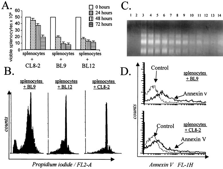
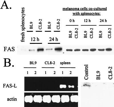
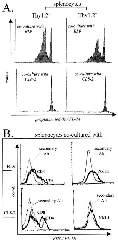
A deranged expression of MHC class I glycoproteins, characteristic of a variety of malignancies, contributes to the ability of cancer to avoid destruction by T cell-mediated immunity. An abrogation of the metastatic capacity of B16 melanoma cells has been achieved by transfecting an MHC class I-encoding vector into class I-deficient B16 melanoma clones [Gorelik, E., Kim, M., Duty, L. & Galili, U. (1993) Clin. Exp. Metastasis 11, 439-452]. We report here that the deranged expression of class I molecules by B16 melanoma cells is more than a mere acquisition of the capacity to escape immune recognition. Namely, cells of the B16 melanoma prompted splenic lymphocytes to commit death after coculture. However, a class I-expressing and nonmetastatic CL8-2 clone was found to be less potent as an inducer of apoptosis than class I-deficient and metastatic BL9 and BL12 clones. Both Thy1.2(+) and Thy1.2(-) splenocytes underwent cell death when exposed to the class I-deficient BL9 clone. A proportion of CD4(+) and CD8(+) cells among splenocytes exposed to the BL9 clone was lower than that observed in a coculture with cells of the CL8-2 clone. Consistently, none of the melanoma clones studied produced a ligand to the FAS receptor (FAS-L). Thus, our results provide evidence that (i) the production of FAS-L may not be the sole mechanism by which malignant cells induce apoptosis in immunocytes, and (ii) absence of MHC class I glycoproteins plays an important role in preventing the elimination of potential effector immunocytes by tumor cells.
Figures
Similar articles
-
Attenuation of the Fas-L independent B16BL6 melanoma lymphocidic capacity by H-2K class I molecules.Immunol Lett. 2005 Sep 15;100(2):146-52. doi: 10.1016/j.imlet.2005.03.016. Immunol Lett. 2005. PMID: 15935480
-
Alterations in the expression of MHC class I glycoproteins by B16BL6 melanoma cells modulate insulin receptor-regulated signal transduction and augment [correction of augments] resistance to apoptosis.J Immunol. 2003 Sep 15;171(6):2945-52. doi: 10.4049/jimmunol.171.6.2945. J Immunol. 2003. PMID: 12960318
-
Dendritic cells charged with apoptotic tumor cells induce long-lived protective CD4+ and CD8+ T cell immunity against B16 melanoma.J Immunol. 2003 Dec 1;171(11):5940-7. doi: 10.4049/jimmunol.171.11.5940. J Immunol. 2003. PMID: 14634105
-
Soluble HLA class I molecules/CD8 ligation trigger apoptosis of CD8+ cells by Fas/Fas-ligand interaction.ScientificWorldJournal. 2002 Feb 12;2:421-3. doi: 10.1100/tsw.2002.122. ScientificWorldJournal. 2002. PMID: 12806026 Free PMC article. Review.
-
Modulation by MHC class I antigens of the biology of melanoma cells. Non-immunological mechanisms.Melanoma Res. 1993 Aug;3(4):285-9. Melanoma Res. 1993. PMID: 8219762 Review.
Cited by
-
Life-extended glycosylated IL-2 promotes Treg induction and suppression of autoimmunity.Sci Rep. 2021 Apr 7;11(1):7676. doi: 10.1038/s41598-021-87102-4. Sci Rep. 2021. PMID: 33828163 Free PMC article.
-
A Potent Inhibitor of Phosphoinositide 3-Kinase (PI3K) and Mitogen Activated Protein (MAP) Kinase Signalling, Quercetin (3, 3', 4', 5, 7-Pentahydroxyflavone) Promotes Cell Death in Ultraviolet (UV)-B-Irradiated B16F10 Melanoma Cells.PLoS One. 2015 Jul 6;10(7):e0131253. doi: 10.1371/journal.pone.0131253. eCollection 2015. PLoS One. 2015. PMID: 26148186 Free PMC article.
-
Synthesis and anti-melanoma effect of 3-O-prenyl glycyrrhetinic acid against B16F10 cells via induction of endoplasmic reticulum stress-mediated autophagy through ERK/AKT signaling pathway.Front Oncol. 2022 Aug 2;12:890299. doi: 10.3389/fonc.2022.890299. eCollection 2022. Front Oncol. 2022. PMID: 35982963 Free PMC article.
References
-
- Townsend A, Bodmer H. Annu Rev Immunol. 1989;7:601–624. - PubMed
-
- De Batselier P, Katzav S, Gorelik E, Feldman M, Segal S. Nature (London) 1980;288:179–181. - PubMed
-
- Katzav S, De Batselier P, Tartakovsky B, Feldman M, Segal S. J Natl Cancer Inst. 1983;71:317–324. - PubMed
-
- Algarra I, Gaforio J J, Cabrera T, Collado A, Garrido F. J Biol Regul Homeost Agents. 1999;13:90–96. - PubMed
Publication types
MeSH terms
Substances
LinkOut - more resources
Full Text Sources
Medical
Research Materials
Miscellaneous
