. 2001 Feb 13;98(4):1757-62.
doi: 10.1073/pnas.98.4.1757.

Distinct role of gp130 activation in promoting self-renewal divisions by mitogenically stimulated murine hematopoietic stem cells

Affiliations

Distinct role of gp130 activation in promoting self-renewal divisions by mitogenically stimulated murine hematopoietic stem cells

J Audet et al. Proc Natl Acad Sci U S A. .

Abstract

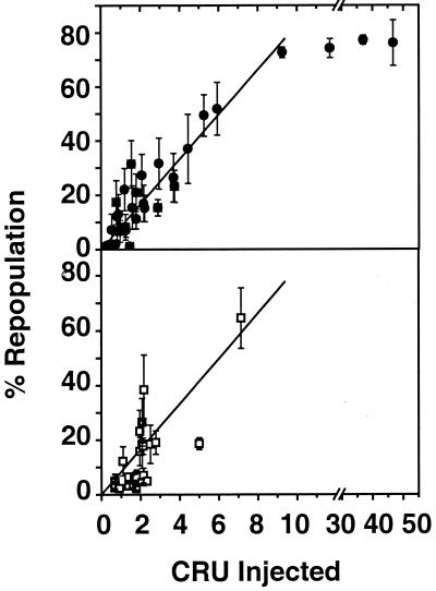
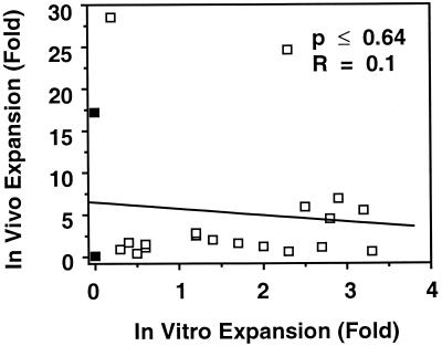
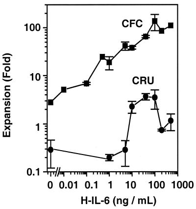
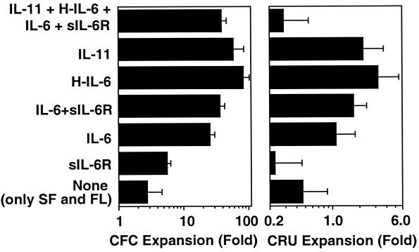
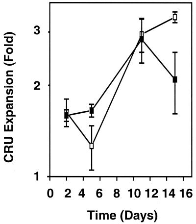
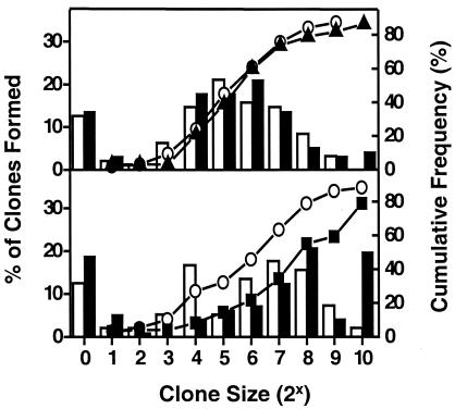
Previous studies have demonstrated hematopoietic stem cell amplification in vitro after the activation of three cell-surface receptors: flt3/flk2, c-kit, and gp130. We now show flt3-ligand and Steel factor alone will stimulate >85% of c-kit(+)Sca-1(+)lin(-) adult mouse bone marrow cells to proliferate in single-cell serum-free cultures, but concomitant retention of their stem cell activity requires additional exposure to a ligand that will activate gp130. Moreover, this response is restricted to a narrow range of gp130-activating ligand concentrations, above and below which hematopoietic stem cell activity is lost. These findings indicate a unique contribution of gp130 signaling to the maintenance of hematopoietic stem cell function when these cells are stimulated to divide with additional differential effects dictated by the intensity of gp130 activation.


Figures

Figure 1
Figure 1
Percent repopulation is linearly related to the number of CRU injected. Percent repopulation is shown as a function of the number of CRU injected into W41 recipients given a sublethal dose of radiation of 400 cGy (●) or B6C3 recipients given a lethal dose of 950 cGy and cotransplanted with 105 syngeneic BM cells (■). Levels of repopulation were assessed 4 mo after the transplant of either fresh (Top) or cultured (Bottom) BM cells from congenic Ly5.1 donors and are expressed as the proportion of lymphoid and myeloid donor-derived (Ly5.1+) cells present in the peripheral blood. The regression line shown in the Top was determined by the method of least squares and has been redrawn in the Bottom to show its closeness of fit to the cultured cell data.
Figure 2
Figure 2
Expansion of CRU in primary recipients is independent of the extent of prior CRU expansion in vitro. Primary recipients of fresh (■) or cultured BM cells (□) containing limiting numbers of CRU were killed 4 mo posttransplant in 21 experiments. Similar numbers of their BM cells were then assayed in secondary recipients to determine the frequency and hence the number of CRU regenerated from the original fresh or cultured CRU injected into the primary recipients. Fold expansions in vivo were calculated by dividing the number of CRU detected 4 mo posttransplant in primary recipients by the corresponding number of CRU calculated to be present initially in the cell suspension injected. Cultured cells were obtained by incubating Sca-1+lin mouse BM cells for 11 days with SF (50 ng/ml) and FL (100 ng/ml) and different combinations and concentrations of gp130-activating cytokines. Fold expansions in vitro were calculated by dividing the number of CRU detected in the 11-day cultures by the corresponding number of CRU calculated to be present in the cells used to initiate each culture. The regression line was obtained by the method of least squares.
Figure 3
Figure 3
Dependence of CRU and CFC expansion in vitro on the concentration of H-IL-6 present. Sca-1+lin mouse BM cells were cultured in serum-free medium containing SF (50 ng/ml) and FL (100 ng/ml) and varying concentrations of H-IL-6. The fold increase in CRU and CFC (±SEM, n = 2–4) was calculated by dividing the number of these cells detected after 10 days by the corresponding numbers present in the number of Sca-1+lin cells used to initiate each culture.
Figure 4
Figure 4
Similar dose effects of different gp130-activating cytokines on CRU and CFC expansion. Sca-1+lin mouse BM cells were cultured in serum-free medium containing SF (50 ng/ml) and FL (100 ng/ml) and various gp130-activating cytokines at the following concentrations: IL-6 at 20 ng/ml, sIL-6R at 400 ng/ml, IL-11 at 100 ng/ml, H-IL-6 at 40 ng/ml when used alone, and H-IL-6 at 10 ng/ml when used in the four-factor combination. The fold increase in CRU and CFC (±SEM, n = 4–8) was calculated by dividing the number of these cells detected after 11 days by the corresponding numbers present in the number of Sca-1+lin cells used to initiate each culture.
Figure 5
Figure 5
The kinetics of CRU expansion in cultures supplemented with IL-11 or H-IL-6 is similar. Changes in CRU numbers are shown as a function of time when Sca-1+lin mouse BM cells were cultured in serum-free medium containing SF at 50 ng/ml and FL at 100 ng/ml and either IL-11 at 100 ng/ml (□) or H-IL-6 at 40 ng/ml (■). The fold increase (±SEM, n = 2–4) was calculated by dividing the number of CRU detected in the cultured populations by the number of CRU present in the number of cells used to initiate each culture.
Figure 6
Figure 6
Presence of gp130-activating cytokines has no effect on the survival or initial mitogenesis c-kit+Sca-1+lin cells also stimulated by SF and FL. Clone size distribution (bars) and cumulative frequency of clones (symbols) generated in 10 days by single PI c-kit+Sca-1+lin mouse BM cells incubated in serum-free medium supplemented with SF (50 ng/ml) and FL (100 ng/ml) in combination with (Top) IL-6 at 50 ng/ml (closed bars and ▴) or no other cytokine (open bars and ○, n = 95 wells); (Bottom) H-IL-6 at 40 ng/ml (closed bars and ■, n = 96 wells) or no other cytokine (open bars and ○, n = 96 wells).

Similar articles

Cited by

References

    1. Yoshida K, Taga T, Saito M, Suematsu S, Kumanogoh A, Tanaka T, Fujiwara H, Hirata M, Yamagami T, Nakahata T, et al. Proc Natl Acad Sci USA. 1996;93:407–411. - PMC - PubMed
    1. Holyoake T, Freshney M, McNair L, Parker A, McKay P, Steward W, Fitzsimons E, Graham G, Pragnell I. Blood. 1996;87:4589–4595. - PubMed
    1. Miller C L, Eaves C J. Proc Natl Acad Sci USA. 1997;94:13648–13653. - PMC - PubMed
    1. Yonemura Y, Ku H, Lyman S D, Ogawa M. Blood. 1997;89:1915–1921. - PubMed
    1. Hirano T, Nakajima K, Hibi M. Cytokine Growth Factor Rev. 1997;8:241–252. - PubMed

Publication types