. 2001 Feb 13;98(4):1763-8.
doi: 10.1073/pnas.98.4.1763.

Identification and modulation of a naturally processed T cell epitope from the diabetes-associated autoantigen human glutamic acid decarboxylase 65 (hGAD65)

Affiliations

Identification and modulation of a naturally processed T cell epitope from the diabetes-associated autoantigen human glutamic acid decarboxylase 65 (hGAD65)

G T Nepom et al. Proc Natl Acad Sci U S A. .

Abstract

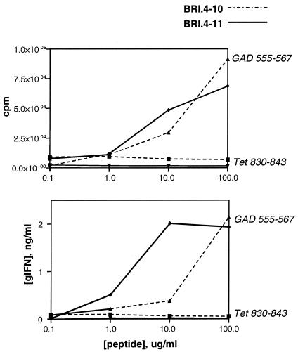
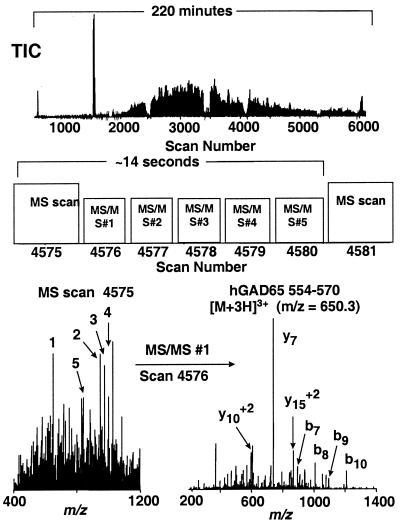
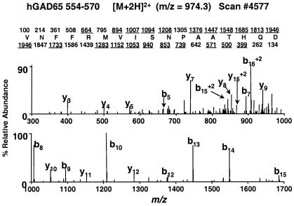
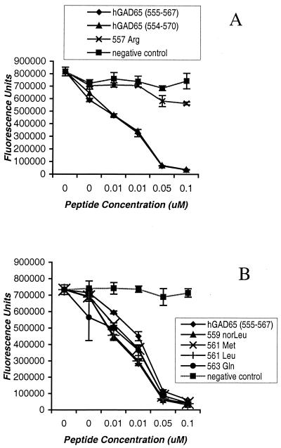
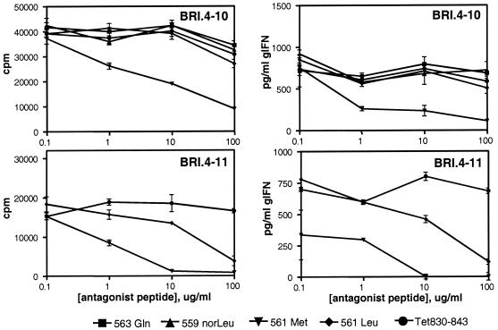
T cell recognition of autoantigens is critical to progressive immune-mediated destruction of islet cells, which leads to autoimmune diabetes. We identified a naturally presented autoantigen from the human islet antigen glutamic acid decarboxylase, 65-kDa isoform (GAD65), by using a combination of chromatography and mass spectrometry of peptides bound by the type I diabetes (insulin-dependent diabetes mellitus, IDDM)-associated HLA-DR4 molecule. Peptides encompassing this epitope-stimulated GAD65-specific T cells from diabetic patients and a DR4-positive individual at high risk for developing IDDM. T cell responses were antagonized by altered peptide ligands containing single amino acid modifications. This direct identification and manipulation of GAD65 epitope recognition provides an approach toward dissection of the complex CD4(+) T cell response in IDDM.


Figures

Figure 1
Figure 1
T cell response profiles for human CD4+ T cell clones BRI.4–10 and BRI.4–11. Proliferation was measured by thymidine uptake (Upper) and IFN-γ release was determined by specific ELISA (Lower). Clones were stimulated with specific GAD peptides or with control peptides derived from tetanus toxoid (Tet 830–843).
Figure 2
Figure 2
Identification of GAD65-derived peptides by using data-dependent MS/MS analysis of an unfractionated HLA-DR4-restricted peptide extract. (Top) Total ion chromatogram (TIC). (Middle) Data acquisition scheme. For MS scan no. 4575 m/z values for all ions in the 300–2,000 range were recorded (MS mode), and the instrument control computer automatically selected the five most abundant ions. Each of these ions was subjected to collision-activated dissociation to yield peptide sequence-specific fragment ions (MS/MS analysis) over the next five scans. The m/z values for these five ions were ignored by the instrument for a time equal to the observed chromatographic peak width (2 min for the data shown herein) to minimize redundant MS/MS analyses (e.g., dissociating the same peptide multiple times). After the next MS scan (no. 4581), the instrument selected the five most abundant ions, exclusive of those already identified in scan no. 4575. In this manner, ions having abundances over a wide dynamic range were automatically subjected to MS/MS analysis. (Bottom Left) MS scan no. 4575, showing ions that were selected for subsequent MS/MS analysis (numbered in order of abundance). (Bottom Right) MS/MS spectrum (scan no. 4576) for m/z = 650.3, the +3 charge state of the hGAD65 peptide 554–570 (corresponding to the first ion selected in MS scan no. 4575). All MS/MS spectra were searched against both an hGAD65 single-protein database and the nonredundant protein database maintained at the National Center for Biotechnology Information by using the SEQUEST search algorithm. The hGAD65 peptide sequence identified by this analysis was confirmed by manual interpretation of the MS/MS spectrum and by comparison with a synthetic peptide.
Figure 3
Figure 3
MS/MS spectrum (scan no. 4578) for m/z = 974.8, the +2 charge state of hGAD65-(554–570). Note that all abundant fragment ions observed correspond to b-type (fragment ions containing the N terminus) and y-type (fragment ions containing the C terminus) sequence-specific ions. Predicted type b- and y-fragment ions, respectively, for hGAD-(554–570) are shown above and below the sequence. Ions observed in the mass spectrum are underlined.
Figure 4
Figure 4
Competitive peptide binding to HLA-DR4 (DRB1*0101/DRB1*0401) molecules. Purified DR4 molecules were incubated with 0.1 μM biotinylated peptide standard (IAFTSEHSHFSLK) in the presence of various concentrations of GAD65 analogs, compared with hGAD65-(555–567) (NFFRMVISNPAAT). A nonbinding peptide standard (KSAVLEGTLTAEK) was used as a negative control.
Figure 5
Figure 5
T cell response to GAD-(555–567) in the presence of altered peptide ligands. Proliferative responses (Left) and INF-γ release (Right) are shown for T cell clones BRI.4–10 (Upper) and BRI.4–11 (Lower).

References

    1. Gianani R, Eisenbarth G S. In: Type I Diabetes. Molecular, Cellular, and Clinical Immunology. Eisenbarth G S, Lafferty K J, editors. New York: Oxford Univ. Press; 1996. pp. 209–229.
    1. Nepom G T. Curr Opin Immunol. 1995;7:825–830. - PubMed
    1. Mehta V, Palmer J P. In: Prediction, Prevention and Genetic Counseling in IDDM. Palmer J P, editor. Chichester, PA: Wiley; 1996. pp. 3–16.
    1. Lernmark A. J Intern Med. 1996;240:259–277. - PubMed
    1. Yoon J W, Yoon C S, Lim H W, Huang Q Q, Kang Y, Pyun K H, Hirasawa K, Sherwin R S, Jun H S. Science. 1999;284:1183–1187. - PubMed

Publication types