. 2001 Feb 13;98(4):1853-8.
doi: 10.1073/pnas.98.4.1853.

Integrin activation controls metastasis in human breast cancer

Affiliations

Integrin activation controls metastasis in human breast cancer

B Felding-Habermann et al. Proc Natl Acad Sci U S A. .

Abstract

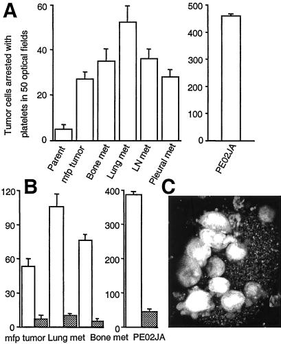
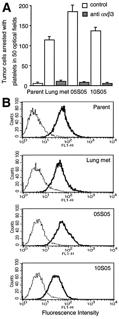
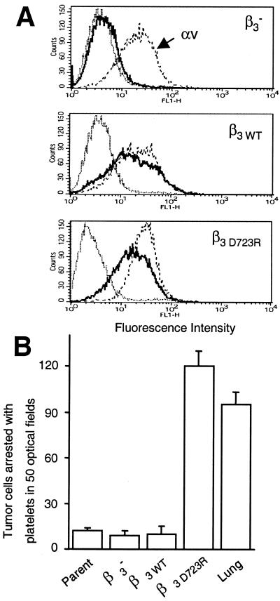
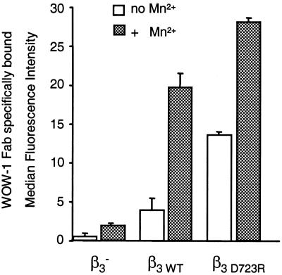
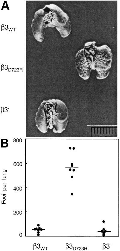
Metastasis is the primary cause of death in human breast cancer. Metastasis to bone, lungs, liver, and brain involves dissemination of breast cancer cells via the bloodstream and requires adhesion within the vasculature. Blood cell adhesion within the vasculature depends on integrins, a family of transmembrane adhesion receptors, and is regulated by integrin activation. Here we show that integrin alpha v beta 3 supports breast cancer cell attachment under blood flow conditions in an activation-dependent manner. Integrin alpha v beta 3 was found in two distinct functional states in human breast cancer cells. The activated, but not the nonactivated, state supported tumor cell arrest during blood flow through interaction with platelets. Importantly, activated alpha v beta 3 was expressed by freshly isolated metastatic human breast cancer cells and variants of the MDA-MB 435 human breast cancer cell line, derived from mammary fat pad tumors or distant metastases in severe combined immunodeficient mice. Expression of constitutively activated mutant alpha v beta 3(D723R), but not alpha v beta 3(WT), in MDA-MB 435 cells strongly promoted metastasis in the mouse model. Thus breast cancer cells can exhibit a platelet-interactive and metastatic phenotype that is controlled by the activation of integrin alpha v beta 3. Consequently, alterations within tumors that lead to the aberrant control of integrin activation are expected to adversely affect the course of human breast cancer.


Figures

Figure 1
Figure 1
Metastatic human breast cancer cells use αvβ3 to interact with platelets and arrest during blood flow. (A) Primary metastatic human breast cancer cells (PE02JA) and MDA-MB 435 cell variants derived from mammary fat pad (mfp) tumors or metastases to bone, lungs, lymph node (LN), or the pleural cavity in mice, but not the parental MDA-MB 435 cell population at large, bind to activated platelets and thereby arrest during blood flow. Tumor cells stained with hydroethidine (red fluorescence) were suspended in human blood (containing mepacrine, green fluorescence) and perfused over a thrombogenic collagen I matrix at a venous wall shear rate (50 s−1, 4 dynes/cm2) (10). Platelets adhere to the matrix, become activated, and form thrombi, to which arrest-competent tumor cells attach (monitored by video microscopy and image acquisition at predefined positions with filter settings to discern platelet- and tumor cell-specific signals). None of the tumor cells attached directly to the matrix. Thrombus formation was not affected by tumor cell type. (B) Arrest-competent breast cancer cells use integrin αvβ3 for attachment. Tumor cells in blood were perfused and analyzed as in A in the absence (open bars) or presence (hatched bars) of function-blocking anti-αvβ3 mAb VNR1 27.1 (18). Columns represent the means of triplicate runs (±SD) with blood from the same donor. (C) Projection of confocal sections through a heteroaggregate of platelets and primary metastatic breast cancer cells, PE02JA (images acquired during perfusion). Attaching to a thrombus, tumor cells extend pseudopods for continued anchorage.
Figure 2
Figure 2
MDA-MB 435 breast cancer cells contain an arrest-competent subset that expresses activated αvβ3. Parental MDA-MB 435 breast cancer cells were suspended in human blood and perfused as in Fig. 1, but under sterile conditions. Arrested cells were expanded and resorted four times. (A) Two independently sorted polyclonal populations (05S05 and 10S05) analyzed for their ability to undergo platelet mediated arrest during blood flow (as in Fig. 1) in the absence (open bars) or presence (hatched bars) of function-blocking anti-integrin αvβ3 mAb VNR1 27.1. Columns represent means of triplicate runs (±SD) with blood from the same donor. (B) Parental MDA-MB 435 cells (Parent) and their in vivo (Lung met) or in vitro (05S05 and 10S05) selected variants express integrin αvβ3 at similar levels. Flow cytometric analysis was carried out on cells stained with mAb LM609 (anti-αvβ3) (——) or isotype control (⋅⋅⋅⋅⋅) and FITC-anti-mouse IgG.
Figure 3
Figure 3
Integrin αvβ3 activation renders breast cancer cells platelet-interactive and arrest-competent. A variant lacking β3 integrin expression (β3) was selected from parental MDA-MB 435 cells by exposure to an anti-β3 saporin conjugate. β3 cells were stably transfected with the β3 wild-type gene (β3WT) or constitutively activated mutant β3D723R. (A) Flow cytometric analysis of αvβ3 expression. Cells stained with anti-αv (mAb AV-8, – – –) (10), anti-αvβ3 (mAb LM609, ——), or isotype control (⋅⋅⋅⋅⋅) and FITC-anti-mouse IgG. (B) Cells expressing constitutively activated αvβ3D723R, but not αvβ3WT, are platelet-interactive and arrest-competent. MDA-MB 435 parental cells (Parent), the β3-lacking variant (β3), transfectants (β3WT, β3D723R), or the in vivo selected metastatic variant (Lung) were perfused in human blood, and cell arrest was analyzed as in Fig. 1. Columns represent means of triplicate runs (±SD) with blood from the same donor.
Figure 4
Figure 4
Binding of the ligand-mimetic antibody Fab WOW-1 to functional variants of MDA-MB 435 breast cancer cells. Flow cytometric analysis of WOW-1 binding (activation-dependent anti-αvβ3 Fab) to variants of MDA-MB 435 breast cancer cells lacking β3 (β3) or transfected with the β3 wild-type gene (β3WT) or constitutively activated mutant β3D723R. Cells were incubated with 10 μg/ml WOW-1 in the absence (open bars) or presence (hatched bars) of 250 μM MnCl2, added to activate αvβ3, and stained with Alexa Fluor 488-anti mouse IgG. Shown is specific WOW-1 binding defined as that inhibited by 2 mM RGDS peptide (means of duplicate analyses ± SD).
Figure 5
Figure 5
Integrin αvβ3 activation controls metastatic potential in the MDA-MB 435 breast carcinoma cell model. (A) Lungs of female C.B17/lcrTac scid mice 42 days after i.v. injection of 1 × 10⁶ tumor cells. The β3 integrin lacking cell variant and its transfectants expressing either αvβ3WT or αvβ3D723R were compared. The β3D723R-expressing variant had the platelet-interactive phenotype and showed increased metastatic activity. (B) Number of metastatic foci at the lung surface. Data points are numbers of lung surface metastases for each animal; horizontal lines are median numbers of metastases per group (n = 8). Cells expressing activated αvβ3D723R produced a significantly larger number of metastases than cells lacking β3 or expressing nonactivated αvβ3WT (P < 0.0001 by the Kruskal–Wallis test).

References

    1. Price J T, Bonovich M T, Kohn E C. Crit Rev Biochem Mol Biol. 1997;32:175–253. - PubMed
    1. Ruoslahti E. Adv Cancer Res. 1999;76:1–20. - PubMed
    1. Schwartz M A, Schaller M D, Ginsberg M H. Annu Rev Cell Dev Biol. 1995;11:549–599. - PubMed
    1. Brooks P C, Clark R A, Cheresh D A. Science. 1994;264:569–571. - PubMed
    1. Albelda S M, Mette S A, Elder D E, Stewart R, Damjanovich L, Herlyn M, Buck C A. Cancer Res. 1990;50:6757–6764. - PubMed

Publication types