. 2001 Feb 13;98(4):1877-82.
doi: 10.1073/pnas.98.4.1877. Epub 2001 Feb 6.

Herpes simplex virus 1 alpha regulatory protein ICP0 functionally interacts with cellular transcription factor BMAL1

Affiliations

Herpes simplex virus 1 alpha regulatory protein ICP0 functionally interacts with cellular transcription factor BMAL1

Y Kawaguchi et al. Proc Natl Acad Sci U S A. .

Abstract

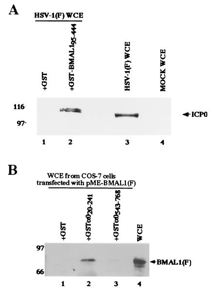
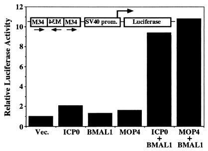
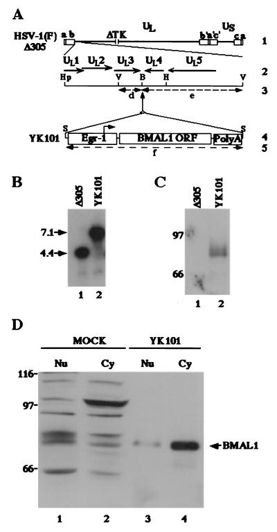
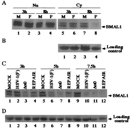
The infected cell protein no. 0 (ICP0) of herpes simplex virus 1 (HSV-1) is a promiscuous transactivator shown to enhance the expression of gene introduced into cells by infection or transfection. At the molecular level, ICP0 is a 775-aa ring finger protein localized initially in the nucleus and late in infection in the cytoplasm and mediates the degradation of several proteins and stabilization of others. None of the known functions at the molecular level account for the apparent activity of ICP0 as a transactivator. Here we report that ICP0 functionally interacts with cellular transcription factor BMAL1, a member of the basic helix-loop-helix PER-ARNT-SIM (PAS) super family of transcriptional regulators. Specifically, sequences mapped to the exon II of ICP0 interacted with BMAL1 in the yeast two-hybrid system and in reciprocal pull-down experiments in vitro. Moreover, the enhancement of transcription of a luciferase reporter construct whose promoter contained multiple BMAL1-binding sites by ICP0 and BMAL1 was significantly greater than that observed by ICP0 or BMAL1 alone. Although the level of BMAL1 present in nuclei of infected cells remained unchanged between 3 and 8 h after infection, the level of cytoplasmic BMAL1 was reduced at 8 h after infection. The reduction of cytoplasmic BMAL1 was significantly greater in cells infected with the ICP0-null mutant than in the wild-type virus-infected cells, suggesting that ICP0 mediates partial stabilization of the protein. These results indicate that ICP0 interacts physically and functionally with at least one cellular transcription-regulatory factor.


Figures

Figure 1
Figure 1
(A) Schematic diagram of the sequence of the HSV-1 genome and the location of the α0 gene. Line 1, a linear representation of the HSV-1 genome. The unique sequences are represented as the regions. The terminal repeats flanking the unique long (UL) and unique short (US) sequences are shown as open rectangles with their designation letters shown above. Line 2, an expanded section of the domain encoding α0 gene. The transcribed and coding domains are shown for one copy of the α0 gene. An identical copy is located in the inverted repeat (b′) flanking UL. Line 3, the region used as “bait” in the yeast two-hybrid screen. Line 4, the domains of the α0 gene fused to GST to generate chimeric proteins.
Figure 2
Figure 2
(A) Photographic image of an immunoblot of infected cell proteins bound to GST or chimeric GST-BMAL1 fusion protein, electrophoretically separated in a denaturing gel and reacted with a mouse mAb to ICP0 (H1083). Lysates of infected HEp-2 were reacted with GST or GST-BMAL1 chimeric protein immobilized on glutathione-agarose beads. The beads were pelleted, rinsed extensively, subjected to electrophoresis on a denaturing gel, transferred to a nitrocellulose sheet, and reacted with the ICP0 antibody. Lines 1 and 2, whole-cell extracts (WCE) from HSV-1(F)-infected HEp-2 cells reacted with GST or GST-BMAL1, respectively; lanes 3 and 4, whole-cell extracts from HSV-1(F)-infected or mock-infected HEp-2 cells, respectively. Molecular weights (×1,000) are shown on the left. (B) Photographic image of an immunoblot of proteins extracted from transfected cells and bound to GCT, GST-α020–241, or GST-α0543–768, electrophoretically separated in a denaturing gel, and reacted with a mouse mAb to the Flag epitope (M2). Lysates of COS-7 cells transfected with pME-BMAL1(F) were reacted with GST or GST-ICP0 chimeric protein immobilized on glutathione-Sepharose beads. The beads were pelleted, rinsed extensively, subjected to electrophoresis on a denaturing gel, transferred to a nitrocellulose sheet, and reacted with the anti-Flag epitope antibody. Lanes 1, 2, and 3, whole-cell extracts from COS-7 cells transfected with pME-BMAL1(F) bound to GST, GST-α020–241, and GST-α0543–768, respectively; lane 4, whole-cell extracts from COS-7 cells transfected with pME-BMAL1(F). Molecular weights (103) are shown on the left.
Figure 3
Figure 3
ICP0-BMAL1 complex enhances transcription of mRNA from the reporter gene containing BMAL1 response elements. COS-7 cells were transfected with the M34 reporter plasmid and pRSV-LacZ in combination with expression plasmid as indicated. Luciferase activity was assayed in cells that were harvested 48 h after transfection as described in Materials and Methods. Luciferase activity were normalized with that of β-galactosidase. The results represent averages of three independent experiments.
Figure 4
Figure 4
(A) Schematic diagram of the sequence of HSV-1(F)Δ305 and the recombinant virus, YK101, derived from it. Line 1, sequence arrangement of the HSV-1(F)Δ305 DNA. Line 2, an enlarged portion of the domain of HSV-1 encoding UL1–UL5. The coding region and direction of the genes are indicated. Line 4, sequence of the recombinant virus, YK101, which was selected, as described in Materials and Methods, from among the progeny of transfection of rabbit skin cells with intact HSV-1(F)Δ305 DNA and plasmid pBMAL1 in UL3–4. The promoter sequence of Egr-1, the BMAL1 coding sequence, and the sequence containing bidirectional poly(A) signals of UL21 and UL22 of HSV-1(F) are shown as open rectangles. The arrow indicates the site of transcription initiation and direction of transcription. Lines 3 and 5 show expected sizes of DNA fragments generated by cleavage of the DNA. The fragment designations shown here are identical to those described in the text and in B. Restriction sites: B, BamHI site; Hp, HpaI site; S, SphI site; and V, EcoRV. (B) Characterization of an HSV-1 recombinant virus expressing the BMAL1 gene (YK101). Autoradiographic image of electrophoretically separated EcoRV digests of HSV-1(F)Δ305 or YK101 DNAs, hybridized to the radiolabeled EcoRV-HindIII fragment of pRB442 that contained sequences of UL3 and UL4. (C) Lane 1, digest of DNA of HSV-1(F)Δ305. Lane 2, digest of DNA of recombinant virus YK101. Molecular weights (×1,000) are shown on the left. (D) Photographic image of an immunoblot of electrophoretically separated lysates of HSV-1(F)Δ305 (lane 1) or YK101 (lane 2) infected with Vero cells harvested 24 h after infection. The blot was probed with the anti-BMAL1 polyclonal antibody. Molecular weights (×1,000) are shown on the left. (D) Photographic image of an immunoblot of electrophoretically separated cell fractions of SK-N-SH cells mock-infected (lanes 1 and 2) or infected with YK101 (lanes 3 and 4). Nuclear (Nu; lanes 1 and 3) and cytoplasmic (Cy; lanes 2 and 4) fractions of infected SK-N-SH cells harvested 24 h after infection were prepared as described in Materials and Methods and reacted with the anti-BMAL1 polyclonal antibody. BMAL1-specific band is indicated on the right, and molecular weights (×1,000) are shown on the left.
Figure 5
Figure 5
(A) Photographic images of electrophoretically separated cell fraction of SK-N-SH cells mock-infected (M) or infected with 10 pfu of HSV-1 (F) per cell and reacted with the rabbit polyclonal antibody to BMAL1. (B) Levels of expression of loading control in the cytoplasmic fractions. Fractions of the infected cells harvested at indicated times were prepared as described in Materials and Methods and reacted with the anti-BMAL1 antibody. (C) Photographic images of electrophoretically separated cytoplasmic fractions of SK-N-SH cells mock-infected or infected with 10 pfu of HSV-1 (F), R7910 (Δa0), or R7911 (REPAIR) per cells and reacted with the polyclonal antibody to BMAL1. (D) The levels of expression of loading control.

References

    1. Ogle W O, Ng T I, Carter K L, Roizman B. Virology. 1997;235:406–413. - PubMed
    1. Purves F C, Roizman B. Proc Natl Acad Sci USA. 1992;89:7310–7314. - PMC - PubMed
    1. Roizman B, Sears A E. In: Fields Virology. 3rd Ed. Fields B N, Knipe D M, Howley P, Chanock R M, Hirsch M S, Melnick J C, Monath J P, Roizman B, editors. Philadelphia: Lippincott; 1996. pp. 2231–2295.
    1. Blaho J A, Mitchell C, Roizman B. J Virol. 1993;67:3891–3900. - PMC - PubMed
    1. Mitchell C, Blaho J A, McCormick A L, Roizman B. J Biol Chem. 1997;272:25394–25400. - PubMed

Publication types

MeSH terms

Substances

LinkOut - more resources