

Neuroimaging of cerebral activations and deactivations associated with hypercapnia and hunger for air
- PMID: 11172070
- PMCID: PMC29376
- DOI: 10.1073/pnas.98.4.2029
Neuroimaging of cerebral activations and deactivations associated with hypercapnia and hunger for air
Abstract
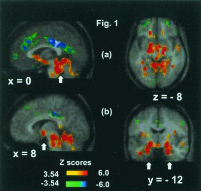
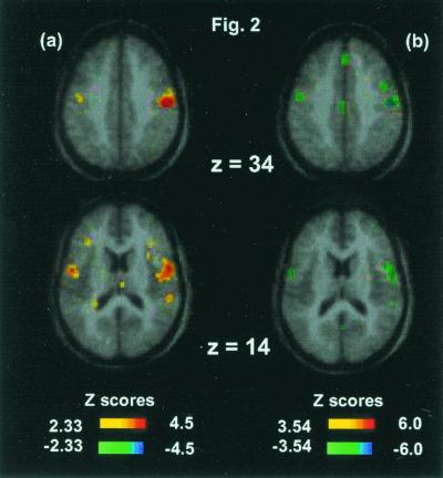
There are defined medullary, mesencephalic, hypothalamic, and thalamic functions in regulation of respiration, but knowledge of cortical control and the elements subserving the consciousness of breathlessness and air hunger is limited. In nine young adults, air hunger was produced acutely by CO(2) inhalation. Comparisons were made with inhalation of a N(2)/O(2) gas mixture with the same apparatus, and also with paced breathing, and with eyes closed rest. A network of activations in pons, midbrain (mesencephalic tegmentum, parabrachial nucleus, and periaqueductal gray), hypothalamus, limbic and paralimbic areas (amygdala and periamygdalar region) cingulate, parahippocampal and fusiform gyrus, and anterior insula were seen along with caudate nuclei and pulvinar activations. Strong deactivations were seen in dorsal cingulate, posterior cingulate, and prefrontal cortex. The striking response of limbic and paralimbic regions points to these structures having a singular role in the affective sequelae entrained by disturbance of basic respiratory control whereby a process of which we are normally unaware becomes a salient element of consciousness. These activations and deactivations include phylogenetically ancient areas of allocortex and transitional cortex that together with the amygdalar/periamygdalar region may subserve functions of emotional representation and regulation of breathing.
Figures
References
Publication types
MeSH terms
Substances
LinkOut - more resources
Full Text Sources
