

Review
doi: 10.1186/gb-2000-1-4-reviews0004.
Epub 2000 Oct 13.
An overview of the potassium channel family
Affiliations
- PMID: 11178249
- PMCID: PMC138870
- DOI: 10.1186/gb-2000-1-4-reviews0004
Item in Clipboard
Review
An overview of the potassium channel family
Genome Biol.
2000.
Abstract
Potassium channels, tetrameric integral membrane proteins that form aqueous pores through which K+ can flow, are found in virtually all organisms; the genomes of humans, Drosophila, and Caenorhabditis elegans contain 30-100 K+ channel genes each. The structure of a bacterial K+ channel, sequence comparisons with other channels and electrophysiological measurements have enabled conclusions about the mechanism of gating and ion flow to be drawn for many other channels.
Figures
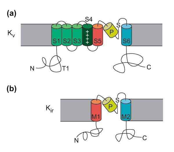
Membrane topologies and main features of the Kv and Kir potassium channel subtypes. (a, b) Schematic representation of the membrane topology of (a) Kv and (b) Kir channels. Note that one subunit of the tetrameric structure is shown (see Figures 3b, 4). Transmembrane helices are numbered S1-S6 in Kv channels and M1 and M2 in Kir channels; P, pore helix; S, signature sequence; N, amino terminus; C, carboxyl terminus; T1, conserved T1 domain (see Figure 4). The extracellular side is towards the top. Adapted with permission from [24].
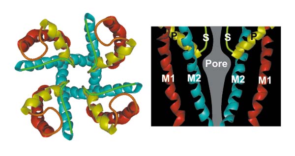
Structure of the Streptomyces lividans K+ channel (KcsA). Labels are as in Figure 1. Reproduced with permission from [24].
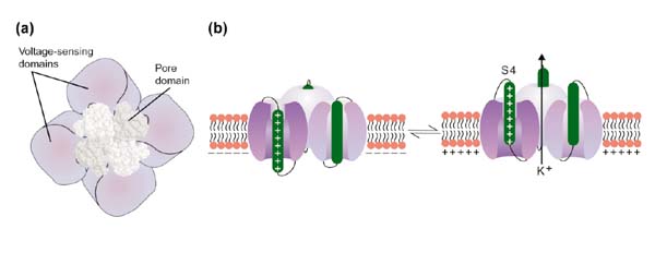
(a) View of a hypothetical Kv-type K+ channel from the extracellular side of the membrane, showing the central pore-forming region, S5-P-S6, modeled by the structure of KcsA, surrounded by a voltage-gating domain of unknown structure, composed of S1-S4. Reproduced with permission from [25]. (b) Diagram of a typical K+ channel, showing the outward movement of the voltage-sensing transmembrane segment S4 concomitant with channel opening. Reproduced with permission from [26].
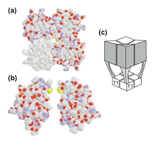
The amino-terminal T1 domain of Kv channels. (a, b) Structure of the soluble, isolated T1 domain of Kv 1.1 from Aplysia californica, with views along (a) the fourfold axis and (b) perpendicular to it. (c) Schematic diagram of the T1 domain location in the full-length K+ channel. Reproduced with permission from [18].
References
-
- Hille B. Ionic Channels of Excitable Membranes. . Sunderland, Massachusetts: Sinauer Associates, 1992.
-
- Littleton JT, Ganetzky B. Ion channels and synaptic organization: analysis of the Drosophila genome. . Neuron. 2000;26:35–43. - PubMed
-
- Papazian DM, Schwarz TL, Tempel BL, Jan YN, Jan LY. Cloning of genomic and complementary DNA from Shaker, a putative potassium channel gene from Drosophila. Science. 1987;237:749–753. - PubMed
-
- Ho K, Nichols CG, Lederer WJ, Lytton J, Vassilev PM, Kanazirska MV, Hebert SC. Cloning and expression of an inwardly rectifying ATP-regulated potassium channel. Nature. 1993;362:31–38. - PubMed
Publication types
MeSH terms
Substances
LinkOut - more resources
Full Text Sources
Other Literature Sources
