

Cytochromes P450: a success story
- PMID: 11178272
- PMCID: PMC138896
- DOI: 10.1186/gb-2000-1-6-reviews3003
Cytochromes P450: a success story
Abstract
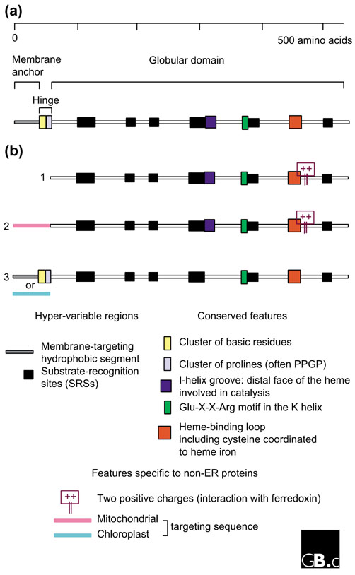
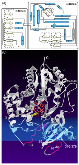
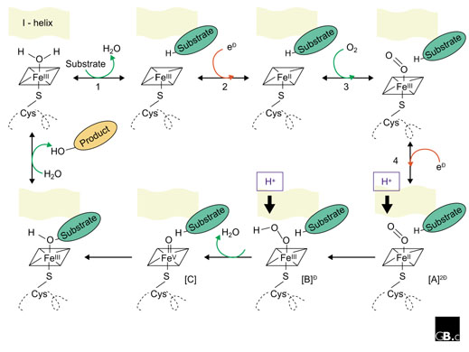
Cytochrome P450 proteins, named for the absorption band at 450 nm of their carbon-monoxide-bound form, are one of the largest superfamilies of enzyme proteins. The P450 genes (also called CYP) are found in the genomes of virtually all organisms, but their number has exploded in plants. Their amino-acid sequences are extremely diverse, with levels of identity as low as 16% in some cases, but their structural fold has remained the same throughout evolution. P450s are heme-thiolate proteins; their most conserved structural features are related to heme binding and common catalytic properties, the major feature being a completely conserved cysteine serving as fifth (axial) ligand to the heme iron. Canonical P450s use electrons from NAD(P)H to catalyze activation of molecular oxygen, leading to regiospecific and stereospecific oxidative attack of a plethora of substrates. The reactions carried out by P450s, though often hydroxylation, can be extremely diverse and sometimes surprising. They contribute to vital processes such as carbon source assimilation, biosynthesis of hormones and of structural components of living organisms, and also carcinogenesis and degradation of xenobiotics. In plants, chemical defense seems to be a major reason for P450 diversification. In prokaryotes, P450s are soluble proteins. In eukaryotes, they are usually bound to the endoplasmic reticulum or inner mitochondrial membranes. The electron carrier proteins used for conveying reducing equivalents from NAD(P)H differ with subcellular localization. P450 enzymes catalyze many reactions that are important in drug metabolism or that have practical applications in industry; their economic impact is therefore considerable.
Figures
References
-
- Nelson DR, Koymans L, Kamataki T, Stegeman JJ, Feyereisen R, Waxman DJ, Waterman MR, Gotoh O, Coon MJ, Estabrook RW, et al. P450 superfamily: update on new sequences, gene mapping, accession numbers and nomenclature. Pharmacogenetics. 1996;6:1–42. This paper defines the rules for P450 gene nomenclature, comments on some general characteristics of the P450 gene superfamilly, and gives a list (with references) of P450 genes identified in various organisms. - PubMed
-
- David Nelson's homepage http://drnelson.utmem.edu/CytochromeP450.html David Nelson keeps an update of P450 sequences characterized in all organisms and is also the 'P450 nomenclature committee', the person to contact to obtain a name for a new P450 sequence. Nelson also provides a simple introductory lecture on mammalian P450 enzymes and on all their physiological and other functions.
-
- Nelson DR. Cytochrome P450 and the individuality of species. Arch Biochem Biophys. 1999;369:1–10. doi: 10.1006/abbi.1999.1352. This paper defines the clans in P450 classification and gives some examples. In addition to evolutionary considerations, it comments on new P450 genes and functions recently discovered and on P450 polymorphism and its impact on drug metabolism. - DOI - PubMed
-
- Ingelman-Sundberg M, Daly AK, Oscarson M, Nebert D. Human cytochrome P450 (CYP) gene: recommandations for the nomenclature of alleles. Pharmacogenetics. 2000;10:91–93. doi: 10.1097/00008571-200002000-00012. This paper and the related website [6] give rules and examples for the nomenclature of P450 alleles. - DOI - PubMed
-
- Home Page of the Human Cytochrome P450 (CYP) Allele Nomenclature Committee http://www.imm.ki.se/CYPalleles/ An update of the knowledge on allelic variants of the human genes; also provides instructions on the submission of new variants.
Publication types
MeSH terms
Substances
LinkOut - more resources
Full Text Sources
Other Literature Sources
