. 2001 Mar;68(3):627-41.
doi: 10.1086/318792.

Analyses of the effects that disease-causing missense mutations have on the structure and function of the winged-helix protein FOXC1

Affiliations

Analyses of the effects that disease-causing missense mutations have on the structure and function of the winged-helix protein FOXC1

R A Saleem et al. Am J Hum Genet. 2001 Mar.

Abstract

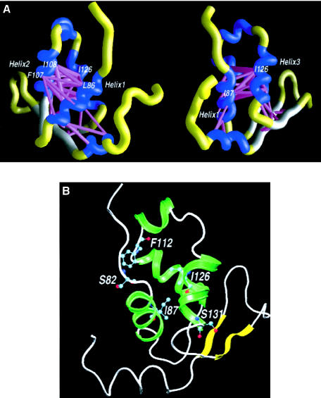
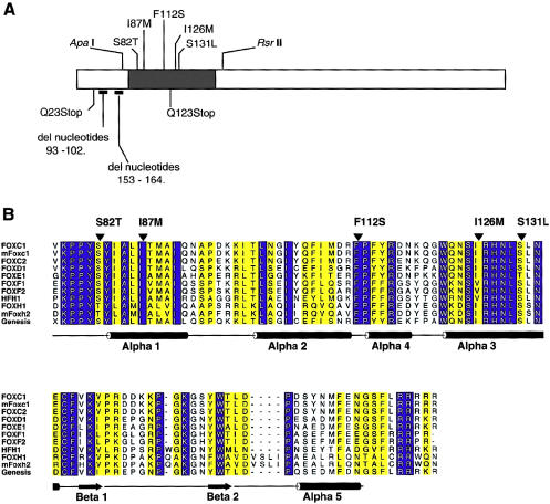
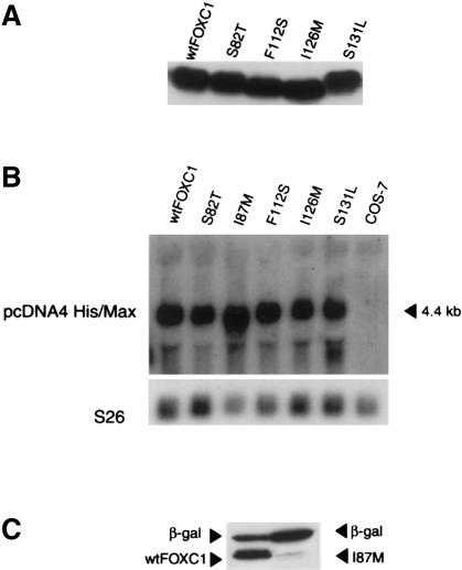
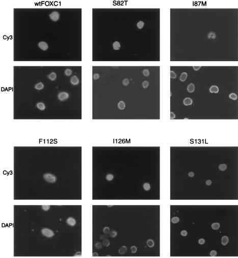
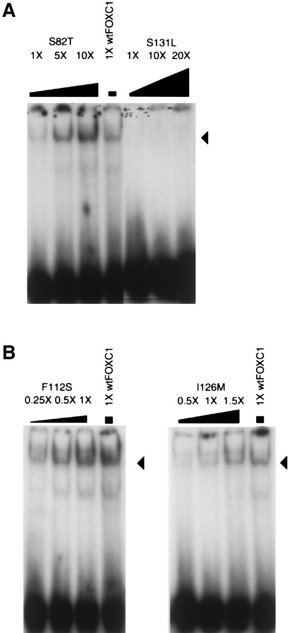
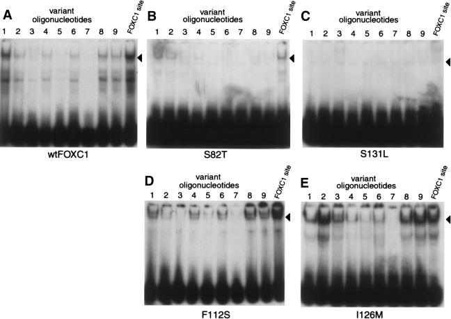
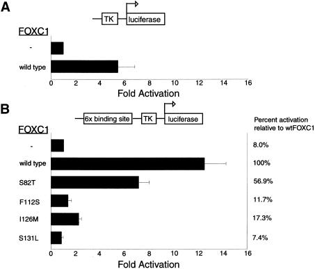
Five missense mutations of the winged-helix FOXC1 transcription factor, found in patients with Axenfeld-Rieger (AR) malformations, were investigated for their effects on FOXC1 structure and function. Molecular modeling of the FOXC1 forkhead domain predicted that the missense mutations did not alter FOXC1 structure. Biochemical analyses indicated that, whereas all mutant proteins correctly localize to the cell nucleus, the I87M mutation reduced FOXC1-protein levels. DNA-binding experiments revealed that, although the S82T and S131L mutations decreased DNA binding, the F112S and I126M mutations did not. However, the F112S and I126M mutations decrease the transactivation ability of FOXC1. All the FOXC1 mutations had the net effect of reducing FOXC1 transactivation ability. These results indicate that the FOXC1 forkhead domain contains separable DNA-binding and transactivation functions. In addition, these findings demonstrate that reduced stability, DNA binding, or transactivation, all causing a decrease in the ability of FOXC1 to transactivate genes, can underlie AR malformations.


Figures

Figure  1
Figure 1
A, Energy scaffolds for the forkhead domain of FOXC1. The α-carbon backbone of the protein is depicted as a curving “worm.” Within the backbone, segments of the forkhead domain that comprise the core folding motifs are shown in blue, whereas the intervening loop regions are shown in yellow. Helices and loop regions are as defined in figure 2B. Pairwise residue-interaction energies between core residues (Bryant and Lawrence 1993) are shown by the width and coloring of the connected α-carbon positions on the protein backbone. Indicated interactions are limited to those with pairwise residue-interaction energies ⩽−1 kcal/mol (“critical” pairwise interactions). Thick, magenta-colored cylinders denote the most favorable interactions; cylinders of intermediate thickness denote interactions with lower, less-favorable pairwise energies. Scaffolds were generated by the graphics program GRASP (Nicholls et al. 1991). The amino acid numbering corresponds to that in the multiple-sequence alignment presented in figure 2B. The second view is 70° counterclockwise around the Y-axis, with respect to the first view. B, Molecular model of the forkhead domain of FOXC1. A ribbon model of the backbone of FOXC1 is shown, with the side chains of the mutated amino acid residues shown in a “ball-and-sticks” representation. The missense mutations in FOXC1 shown here are S82T at the N-terminal end of helix 1, I87M in helix 1, F112S at the C-terminal end of helix 2, and I126M and S131L in helix 3. The numbering in the figure corresponds to that of the human FOXC1 sequence.
Figure  2
Figure 2
A, Schematic of the FOXC1 protein. The blackened rectangle represents the forkhead domain, and the missense mutations studied are indicated above the forkhead domain. Mutations presented below the ideogram result in truncated products and were not tested. Positions of restriction-enzyme sites used in subcloning are indicated above the ideogram. B, Multiple-sequence alignment of the forkhead domain of human FOXC1 and related FOX proteins. The sequences shown in single-letter amino acid codes are those of human FOXC1 (AF048693), mouse Foxc1 (NM008592), human FOXC2 (Y08223), human FOXD1 (U13222), human FOXE1 (U89995), human FOXF1 (U13219), human FOXF2 (U13220), human HFH1 (AF153341), human FOXH1 (AF076292), mouse Foxh2 (AF110506), and rat Genesis/Foxd3 (NM012183), respectively (NCBI Databases) . Amino acid residues showing absolute identity among these proteins are shown in white against a blue background; those positions with conservative substitutions are shown against a yellow background. The positions of the α-helices and β-strands, as defined in the NMR structure of Genesis, are schematically represented below the alignment. Positions of mutations and corresponding amino acid changes in FOXC1 are indicated by the blackened arrowheads above the alignment. ALSCRIPT was used to format the alignment.
Figure  3
Figure 3
A, Western blot analysis of both recombinant FOXC1 and FOXC1 containing missense mutations. Transfected COS-7 whole-cell extracts were resolved by SDS-PAGE and by the N-terminal Xpress epitope detected by immunoblotting with a mouse anti-Xpress monoclonal antibody. B, Northern analysis of COS-7 extracts transfected with FOXC1 pcDNA4 His/Max wild-type and missense mutation constructs. [32P]-labeled EcoRI-linearized pcDNA4 His/Max vector is hybridized to the Xpress epitope, detecting an appropriately sized product at 4.4 kb. S26 cDNA was used as a loading control. C, FOXC1 I87M, which reduces FOXC1 levels. COS-7 cells were transiently cotransfected with LacZ pcDNA4 His/Max and either FOXC1 pcDNA4 His/Max or FOXC1 I87M pcDNA4 His/Max. Transfected COS-7 whole-cell extracts were resolved by SDS-PAGE and by the N-terminal Xpress epitope detected by immunoblotting. Only small amounts (<5% of wild type) of the FOXC1 I87M mutant protein were detected.
Figure  4
Figure 4
Immunofluorescence of Xpress epitope–tagged FOXC1 proteins. COS-7 cells were transiently transfected with the FOXC1 pcDNA4 His/Max constructs. In the upper six panels, the Xpress epitope–tagged recombinant FOXC1 proteins are localized to the nucleus, indicated by Cy3 fluorescence. The lower six panels indicate the position of the nuclei, by staining with DAPI. Note that the Cy3 fluorescence in the I87M transfection is weaker than it is in the other mutations.
Figure  5
Figure 5
FOXC1 missense mutations that affect the DNA-binding ability of FOXC1. EMSA using the [32P]-labeled FOXC1 site was incubated with FOXC1 wild-type and missense mutations containing COS-7 protein extracts. Recombinant FOXC1 missense proteins were equalized to recombinant wild-type FOXC1 by western blotting. The position of the FOXC1-DNA complex is indicated by the blackened arrowheads. A, FOXC1 containing the S82T missense mutation, showing reduced affinity for the FOXC1 site. FOXC1 containing the S131L missense mutations shows an ∼20-fold reduction in affinity for the FOXC1 site. B, FOXC1 containing the F112S and I126M mutations, showing DNA binding that is at wild-type or near–wild-type FOXC1-protein levels.
Figure  6
Figure 6
Effect of FOXC1 missense mutations on DNA-binding specificity. EMSAs were performed with [32P]-labeled variant sites (see table 2) incubated with COS-7 protein extracts containing either wild-type or mutant FOXC1. Recombinant proteins were equalized to wild-type FOXC1 by western blotting. The position of the FOXC1-DNA complex is indicated by the blackened arrowheads. The FOXC1 I126M protein shows an increased affinity for variant oligonucleotides 2, 3, 8, and 9, compared with wild-type FOXC1’s affinity for these oligonucleotides. Also, compared with its affinity for the FOXC1 site, I126M shows an increased affinity for variant oligonucleotides 2 and 9.
Figure  7
Figure 7
Disruption of the transactivation of a luciferase reporter construct, by FOXC1 missense mutations. The thick blackened bars represent the mean of the values, and the Y error bar represents the SD of the values. In each panel, the upper bar, denoted by a minus sign (−), represents a vector-only control. In panel A, a schematic of the TK promoter–luciferase gene reporter construct is shown above the histogram; transfected HeLa cells were assayed for luciferase activity, as described in the Patients, Material, and Methods section. In panel B, a schematic of the FOXC1 site-TK promoter-luciferase gene reporter construct is shown above the histogram.

References

Electronic-Database Information

    1. National Center for Biotechnology Information (NCBI) Databases, http://www.ncbi.nlm.nih.gov/Database/index.html (for human FOXC1 [AF048693], mouse Foxc1 [NM008592], human FOXC2 [Y08223], human FOXD1 [U13222], human FOXE1 [U89995], human FOXF1 [U13219], human FOXF2 [U13220], human HFH1 [AF153341], human FOXH1 [AF076292], mouse Foxh2 [AF110506], and rat Genesis/Foxd3 [NM012183])
    1. Online Mendelian Inheritance in Man (OMIM), http://www.ncbi.nlm.nih.gov/Omim/ (for FOXC1 [MIM 601090], PITX2 [MIM 601542], Oct1 [MIM 602607], and FOXC2 [MIM 602402])

References

    1. Alward WL, Semina EV, Kalenak JW, Heon E, Sheth BP, Stone EM, Murray JC (1998) Autosomal dominant iris hypoplasia is caused by a mutation in the Rieger syndrome (RIEG/PITX2) gene. Am J Ophthalmol 125:98–100 - PubMed
    1. Becker R, Chambers J, Wilks A (1988) The new S language—a programming environment for data analysis and graphics. Wadsworth, Pacific Grove, CA
    1. Blixt A, Mahlapuu M, Aitola M, Pelto-Huikko M, Enerback S, Carlsson P (2000) A forkhead gene, FoxE3, is essential for lens epithelial proliferation and closure of the lens vesicle. Genes Dev 14:245–254 - PMC - PubMed
    1. Bryant SH, Lawrence CE (1993) An empirical energy function for threading protein sequence through the folding motif. Proteins 16:92–112 - PubMed
    1. Fetrow J, Bryant SH (1993) New programs for protein tertiary structure prediction. Biotechnology 11:479–484 - PubMed

Publication types

MeSH terms