. 2001 Feb 15;20(4):650-60.
doi: 10.1093/emboj/20.4.650.

Crystal structure of dihydropyrimidine dehydrogenase, a major determinant of the pharmacokinetics of the anti-cancer drug 5-fluorouracil

Affiliations

Crystal structure of dihydropyrimidine dehydrogenase, a major determinant of the pharmacokinetics of the anti-cancer drug 5-fluorouracil

D Dobritzsch et al. EMBO J. .

Abstract

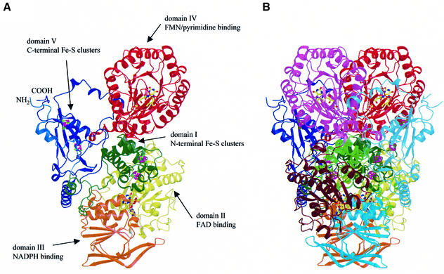
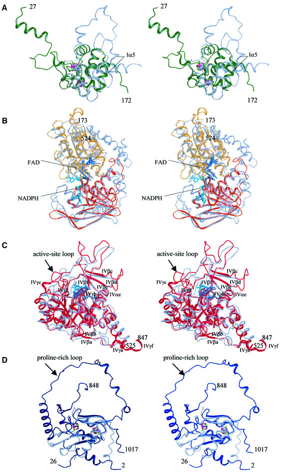
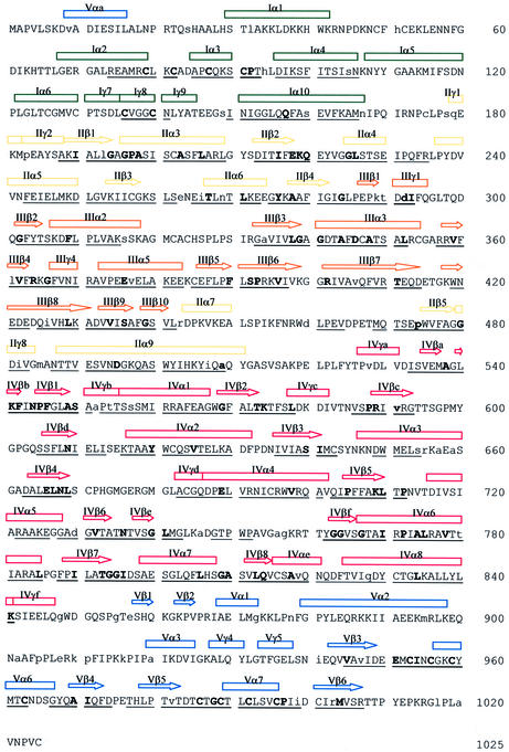
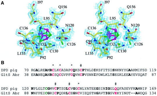
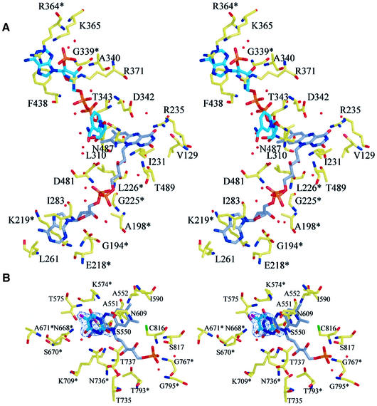
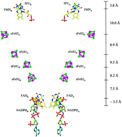
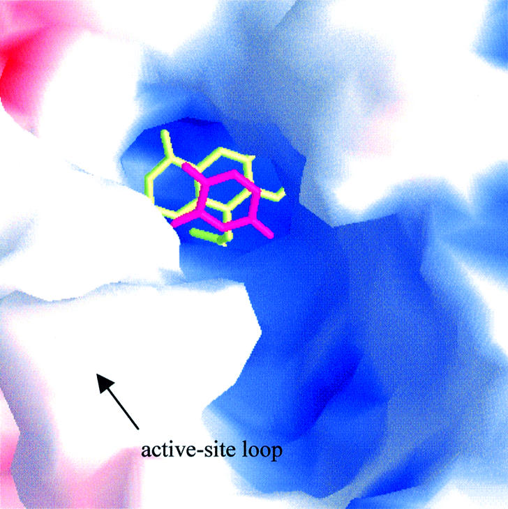
Dihydropyrimidine dehydrogenase catalyzes the first step in pyrimidine degradation: the NADPH-dependent reduction of uracil and thymine to the corresponding 5,6-dihydropyrimidines. Its controlled inhibition has become an adjunct target for cancer therapy, since the enzyme is also responsible for the rapid breakdown of the chemotherapeutic drug 5-fluorouracil. The crystal structure of the homodimeric pig liver enzyme (2x 111 kDa) determined at 1.9 A resolution reveals a highly modular subunit organization, consisting of five domains with different folds. Dihydropyrimidine dehydrogenase contains two FAD, two FMN and eight [4Fe-4S] clusters, arranged in two electron transfer chains that pass the dimer interface twice. Two of the Fe-S clusters show a hitherto unobserved coordination involving a glutamine residue. The ternary complex of an inactive mutant of the enzyme with bound NADPH and 5-fluorouracil reveals the architecture of the substrate-binding sites and residues responsible for recognition and binding of the drug.


Figures

None
Fig. 1. Structure of pig liver DPD. (A) Schematic view of the subunit of DPD with the domains in different colors. The cofactors are shown as ball-and-stick models, iron ions in magenta and sulfur atoms in green. (B) The DPD dimer. The color codes for the domains of the first subunit are the same as in (A), the corresponding domains in the second subunit are shown in light green, brown, cyan, pink and light blue.
None
Fig. 2. Structure of the domains in DPD. (A) Domain I (green). Superimposed is the second domain of the E.coli fumarate reductase iron-protein (FR-IP, gray). The iron and sulfur atoms in DPD are shown in magenta and green, those of FR-IP in medium and dark gray. (B) Domains II (yellow) and III (orange). Superimposed is the structure of the Bos taurus adrenodoxin reductase (gray). For DPD, FAD and NADPH are shown in blue and cyan, those bound to adrenodoxin reductase in medium and dark gray, respectively. (C) Domain IV (red) superimposed on the structure of L.lactis dihydroorotate dehydrogenase (gray). Except for the barrel components, all secondary structure elements are labeled. For DPD, the cofactor FMN is shown in blue and the anti-cancer drug 5FU in cyan. FMN bound to DHOD(A) is colored dark-gray, the reaction product orotate is shown in medium gray. (D) Domain V (blue), superimposed on domain 5 of Desulfovibrio africanus pyruvate:ferredoxin oxidoreductase (gray). The iron and sulfur atoms in DPD are shown in magenta and green, those of pyruvate:ferredoxin oxidoreductase in medium and dark gray.
None
Fig. 3. Amino acid sequence of pig liver DPD. The secondary structure elements are indicated by arrows for β-strands and cylinders for α- and 310(γ)-helices, respectively. The colour code is as in Figure 1, i.e. green for domain I, yellow for domain II, orange for domain III, red for domain IV and blue for domain V. Underlined letters indicate structural similarities between domain I and the second domain of E.coli FR-IP, domains II and III and B.taurus adrenodoxin reductase, domain IV and L.lactis DHOD(A), and domain V and domain 5 of D.africanus pyruvate:ferredoxin oxidoreductase, respectively. Bold letters indicate sequence and structure conservation between DPD and these enzymes, lower case letters indicate differences between pig liver and human DPD.
None
Fig. 4. Unusual coordination of an Fe–S cluster in DPD. (A) Stereoview of the electron density map contoured around the Fe–S cluster with Q156 as iron ligand. Sulfur atoms are green, iron atoms magenta. (B) Sequence comparison of DPD domain I and the corresponding sequence of Amazona brasiliensis glutamate synthase β-subunit (numbering of the amino acids is according to the SwissProt database). Cluster coordinating residues are shown in magenta, the previously suggested cluster ligand C126 in green. Conserved residues are indicated in bold and asterisks denote the residues coordinating the iron ions of the first Fe–S cluster and # the residues coordinating the second Fe–S cluster.
None
Fig. 5. Ligand-binding sites. (A) Stereoview of the binding site for FAD and NADPH. The carbon atoms of the cofactor FAD are shown in gray, those of the cosubstrate NADPH in cyan. Side chains that are conserved between pig liver DPD and B.taurus adrenodoxin reductase are marked with an asterisk. (B) Stereoview of the substrate-binding site shows 5FU bound adjacent to the cofactor FMN in the C671A mutant. A 2|Fo| – |Fc| map is contoured at 1.2σ for 5FU. Side chains that are conserved between pig liver DPD and L.lactis DHOD(A) are marked with an asterisk.
None
Fig. 6. Electron transfer pathways in DPD. Distances between closest atoms of the cofactors are indicated. The nicotinamide ring of NADPH is shown at its assumed position during electron transfer.
None
Fig. 7. Solvent accessibility of the pyrimidine-binding site. Molecular surface representation of the active site with bound FMN (yellow) and 5FU (magenta) as stick models. The surface is colored according to the electrostatic potential, as calculated with GRASP (Nicholls et al., 1991).

References

    1. Ahmed F.Y. et al. (1999) Eniluracil treatment completely inactivates dihydropyrimidine dehydrogenase in colorectal tumors. J. Clin. Oncol., 17, 2439–2445. - PubMed
    1. Albin N., Johnson,M.R. and Diasio,R.B. (1996) cDNA cloning of bovine liver dihydropyrimidine dehydrogenase. DNA Seq., 6, 243–250. - PubMed
    1. Brito R.A., Medgyesy,D., Zukowski,T.H., Royce,M.E., Ravandi-Kashani,F., Hoff,P.M. and Pazdur,R. (1999) Fluoropyrimidines: a critical evaluation. Oncology, 57 (suppl. 1), 2–8. - PubMed
    1. Brünger A.T. et al. (1998) Crystallography & NMR system: a new software suite for macromolecular structure determination. Acta Crystallogr. D, 54, 905–921. - PubMed
    1. Bruschi M. and Guerlesquin,F. (1988) Structure, function and evolution of bacterial ferredoxins. FEMS Microbiol. Rev., 54, 155–176. - PubMed

Publication types