. 2001 Feb 15;20(4):672-82.
doi: 10.1093/emboj/20.4.672.

Vascular endothelial growth factor-C-mediated lymphangiogenesis promotes tumour metastasis

Affiliations

Vascular endothelial growth factor-C-mediated lymphangiogenesis promotes tumour metastasis

S J Mandriota et al. EMBO J. .

Abstract

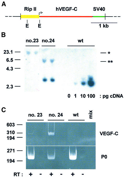
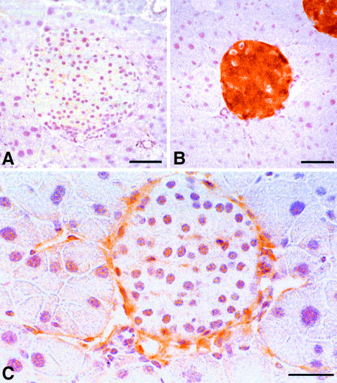
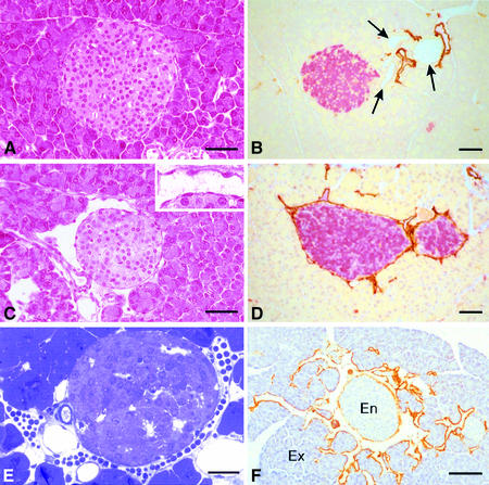
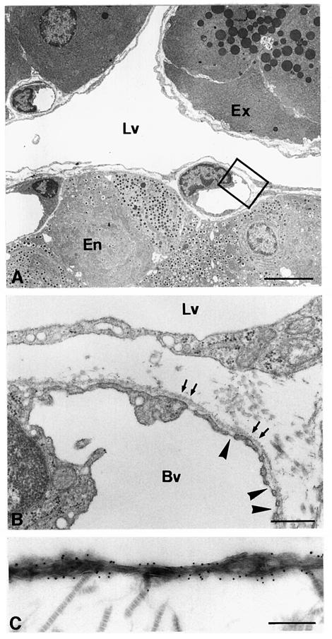
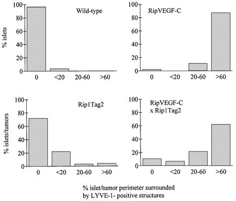
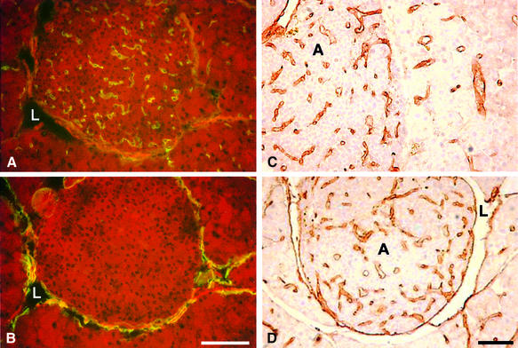
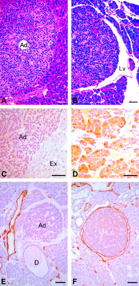
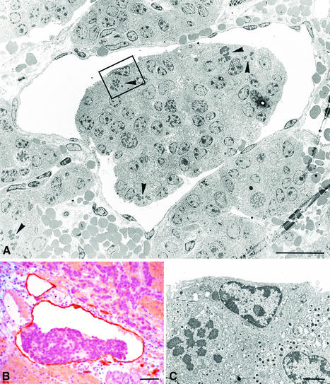
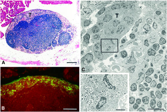
Metastasis is a frequent and lethal complication of cancer. Vascular endothelial growth factor-C (VEGF-C) is a recently described lymphangiogenic factor. Increased expression of VEGF-C in primary tumours correlates with dissemination of tumour cells to regional lymph nodes. However, a direct role for VEGF-C in tumour lymphangiogenesis and subsequent metastasis has yet to be demonstrated. Here we report the establishment of transgenic mice in which VEGF-C expression, driven by the rat insulin promoter (Rip), is targeted to beta-cells of the endocrine pancreas. In contrast to wild-type mice, which lack peri-insular lymphatics, RipVEGF-C transgenics develop an extensive network of lymphatics around the islets of Langerhans. These mice were crossed with Rip1Tag2 mice, which develop pancreatic beta-cell tumours that are neither lymphangiogenic nor metastatic. Double-transgenic mice formed tumours surrounded by well developed lymphatics, which frequently contained tumour cell masses of beta-cell origin. These mice frequently developed pancreatic lymph node metastases. Our findings demonstrate that VEGF-C-induced lymphangiogenesis mediates tumour cell dissemination and the formation of lymph node metastases.


Figures

None
Fig. 1. Molecular characterization of RipVEGF-C transgenic mice. (A) The transgene was constructed by cloning the complete human VEGF-C cDNA (nucleotides 1–1997; DDBJ/EMBL/GenBank accession No. X94216) between the ∼695 bp BamHI–XbaI fragment of Rip (Hanahan, 1985) and the SV40 small T antigen intron and polyadenylation signal. The L-shaped arrow indicates insulin gene transcription initiation. E, EcoRI restriction sites. (B) Ten micrograms of genomic DNA from two RipVEGF-C transgenic mice from families 23 and 24 or from a wild-type littermate (wt) (the latter spiked with the RipVEGF-C transcriptional unit as indicated in picograms) were digested with EcoRI and analysed by Southern blotting using the SV40 moiety of the transgene as a probe. Single or double asterisks indicate the 3′ end of the insertion site in family 23 and 24, respectively. Markers on the left indicate kilobases. (C) Reverse transcription products from oligo dT-primed total RNAs from pancreata of RipVEGF-C transgenic mice from families 23 and 24 or from wild-type mice (wt) were analysed by PCR using hVEGF-C specific primers or acidic ribosomal phosphoprotein P0 (P0) primers. Where indicated, RT was omitted, or PCR mix alone (mix) was analysed. Markers on the left indicate base pairs.
None
Fig. 2. Expression of VEGF-C and VEGFR-3 in RipVEGF-C transgenic mice. Immunohistochemistry for (A and B) VEGF-C and (C) VEGFR-3. (A) Ten-month-old female wild-type-littermate; (B and C) 11-month-old female RipVEGF-C mouse, family 24. (B) and (C) are serial sections. Bar: (A and B) 50 µm; (C) 30 µm.
None
Fig. 3. Histological analysis of wild-type and RipVEGF-C mice. (A and C) Haematoxylin and eosin staining; (B, D and F) immunohistochemistry for LYVE-1 (DAB brown) and insulin (B and D) (fast red); (E) semithin section stained with methylene blue. (A) Twelve-month-old male wild-type littermate; (B) 2-month-old male wild-type littermate; (C) 12-month-old male RipVEGF-C mouse, family 24; (D) 2-month-old male RipVEGF-C mouse, family 24; (E) 11-month-old male RipVEGF-C mouse, family 24; (F) 10-month-old male RipVEGF-C mouse, family 24. Arrows in (B) indicate ducts; En, endocrine; Ex, exocrine. Bar: (A–D) 50 µm; (E) 25 µm; (F) 100 µm.
None
Fig. 4. Ultrastructural analysis of RipVEGF-C transgenic mouse pancreas. (A and B) TEM of the pancreas from a 2-month-old male RipVEGF-C transgenic mouse from family 24. (B) is a higher magnification of the area delimited in (A). Arrows in (B) indicate the basement membrane surrounding a capillary blood vessel (Bv). Note the absence of a basement membrane along the basal surface of the lymphatic endothelium. Arrowheads indicate endothelial fenestrations in the capillary blood vessel. Note the absence of fenestrations in the lymphatic endothelium. (C) Immunoelectron microscopy analysis of LYVE-1 distribution on lymphatic endothelium in the same animal as shown in (A) and (B). Cross-striated collagen fibrils are on the abluminal side of the vessel. Lv, lymphatic vessel; Ex, exocrine tissue; En, endocrine tissue. Bar: (A) 5 µm; (B) 500 nm; (C) 250 nm.
None
Fig. 5. Quantitation of lymphatic vessel density and disposition in wild-type, single- (RipVEGF-C and Rip1Tag2) and double- (RipVEGF-C/Rip1Tag2) transgenic mice. Lymphatic vessels were identified by LYVE-1 immunoreactivity, and the per cent islet/insulinoma perimeter surrounded by LYVE-1-positive structures was determined. For wild type, 185 islets were analysed from five mice; for RipVEGF-C, 99 islets were analysed from five mice; for Rip1Tag2, 232 islets were analysed from seven mice; for RipVEGF-C/Rip1Tag2, 322 islets were analysed from eight mice.
None
Fig. 6. MECA-32 and CD31 immunohistochemistry in wild-type and transgenic islets. (A) MECA-32 and (B) LYVE-1 immunolabelling in consecutive frozen sections of a RipVEGF-C islet. CD31 immunolabelling of adenomas from 14-week-old (C) Rip1Tag2 and (D) double-transgenic RipVEGF-C × Rip1Tag2 mice. L, lymphatic vessel; A, adenoma. Bar: 100 µm.
None
Fig. 7. Histological analysis of RipVEGF-C/Rip1Tag2 double-transgenic mice. (A and B) Haematoxylin and eosin staining; (C and D) anti-VEGF-C immunohistochemistry; (E and F) anti-LYVE-1 immunohistochemistry. (A, C and E) Rip1Tag2 single-transgenic mice. (B, D and F) RipVEGF-C/Rip1Tag2 double-transgenic mice. Ad, adenoma; Lv, lymphatic vessel; Ex, exocrine tissue; D, duct. The white space around the adenoma in (E) is artefactual. All mice were killed at 14 weeks; double-transgenic mice are from RipVEGF-C family 23. Bar: 50 µm.
None
Fig. 8. Intra-lymphatic tumour cell masses in RipVEGF-C/Rip1Tag2 double-transgenic mice. (A) TEM showing a tumour cell mass within an endothelial-lined lymphatic space. (B) Anti-LYVE-1 immunohistochemistry. (C) Enlargement of the area delimited in (A) showing a mitotic figure and endocrine secretory granules. Mice were killed at 14 weeks and were generated from RipVEGF-C family 23. Arrowheads in (A) indicate mitotic figures. Bars: (A) 20 µm; (B) 50 µm; (C) 2 µm.
None
Fig. 9. Lymph node metastases in RipVEGF-C/Rip1Tag2 double-transgenic mice. (A) Haematoxylin and eosin staining of a lymph node containing a peripheral ring of tumour cells (T). (B) Immunofluorescent antibody staining for insulin. (C) TEM showing tumour cells (T) surrounded by lymphocytes. The inset is an enlargement of the delimited area, and shows the presence of endocrine secretory granules in tumour cells. Mice were killed at 14 weeks and were generated from RipVEGF-C family 23. Bar: (A) 200 µm; (B) 50 µm; (C) 10 µm; inset = 2 µm.

Similar articles

Cited by

References

    1. Akagi K., Ikeda,Y., Miyazaki,M., Abe,T., Kinoshita,J., Maehara,Y. and Sugimachi,K. (2000) Vascular endothelial growth factor-C (VEGF-C) expression in human colorectal cancer tissues. Br. J. Cancer, 83, 887–891. - PMC - PubMed
    1. Alpert S., Hanahan,D. and Teitelman,G. (1988) Hybrid insulin genes reveal a developmental lineage for pancreatic endocrine cells and imply a relationship with neurons. Cell, 53, 295–308. - PubMed
    1. Banerji S., Ni,J., Wang,S.X., Clasper,S., Su,J., Tammi,R., Jones,M. and Jackson,D.G. (1999) LYVE-1, a new homologue of the CD44 glycoprotein, is a lymph-specific receptor for hyaluronan. J. Cell Biol., 144, 789–801. - PMC - PubMed
    1. Bertelli E., Regoli,M. and Comparini,L. (1993) Histotopographic and ultrastructural study on the lymphatic network of the pancreas in the guinea pig. Acta Anat. (Basel), 147, 233–239. - PubMed
    1. Breiteneder-Geleff S. et al. (1999) Angiosarcomas express mixed endothelial phenotypes of blood and lymphatic capillaries: podoplanin as a specific marker for lymphatic endothelium. Am. J. Pathol., 154, 385–394. - PMC - PubMed

Publication types

Substances