. 2001 Feb 15;20(4):683-93.
doi: 10.1093/emboj/20.4.683.

Rab-interacting lysosomal protein (RILP): the Rab7 effector required for transport to lysosomes

Affiliations

Rab-interacting lysosomal protein (RILP): the Rab7 effector required for transport to lysosomes

G Cantalupo et al. EMBO J. .

Abstract

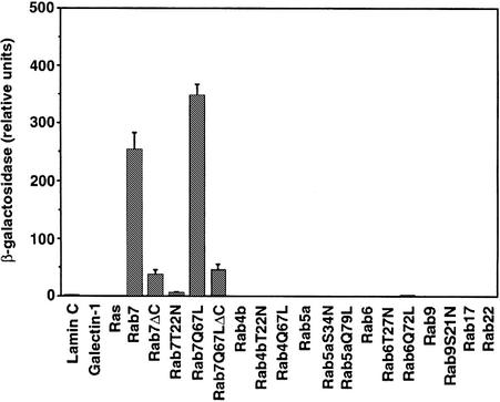
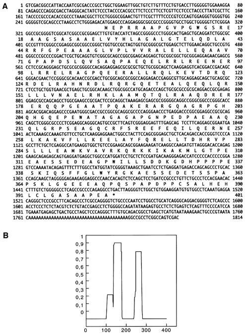
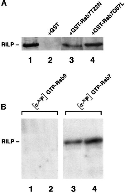
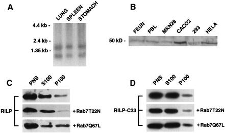
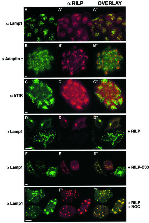
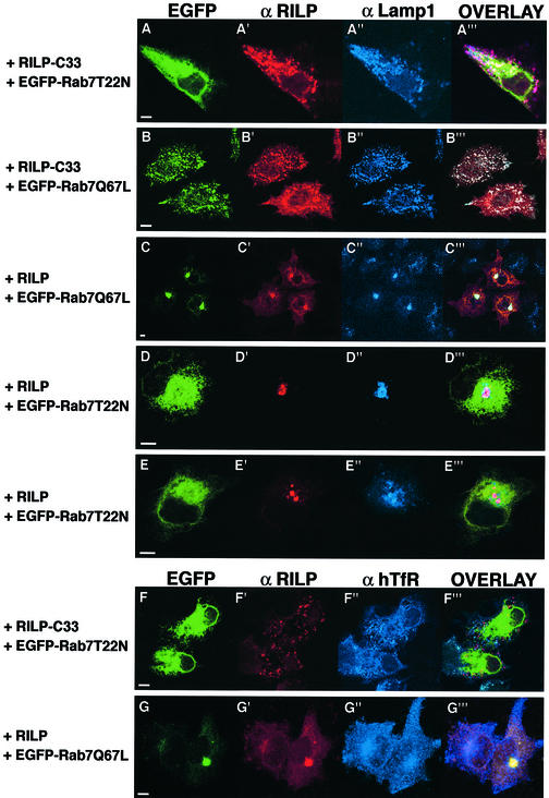
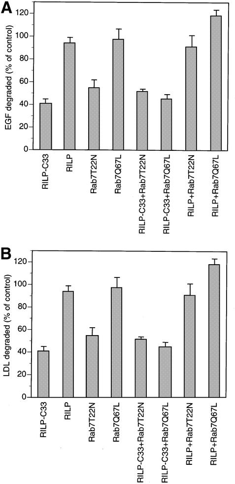
Rab7 is a small GTPase that controls transport to endocytic degradative compartments. Here we report the identification of a novel 45 kDa protein that specifically binds Rab7GTP at its C-terminus. This protein contains a domain comprising two coiled-coil regions typical of myosin-like proteins and is found mainly in the cytosol. We named it RILP (Rab-interacting lysosomal protein) since it can be recruited efficiently on late endosomal and lysosomal membranes by Rab7GTP. RILP-C33 (a truncated form of the protein lacking the N-terminal half) strongly inhibits epidermal growth factor and low-density lipoprotein degradation, and causes dispersion of lysosomes similarly to Rab7 dominant-negative mutants. More importantly, expression of RILP reverses/prevents the effects of Rab7 dominant-negative mutants. All these data are consistent with a model in which RILP represents a downstream effector for Rab7 and both proteins act together in the regulation of late endocytic traffic.


Figures

None
Fig. 1. Specific interaction between Rab7 and C33. The β-galactosidase activity of double transformants was measured using o-nitrophenyl-β-d-galactoside as substrate. Activities are measured as arbitrary relative units and represent mean values ± SEM of four independent transformants.
None
Fig. 2. Primary structure of RILP. (A) Nucleotide and deduced amino acid sequence of RILP. The predicted amino acid sequence is shown below the nucleotide sequence in single-letter code. (B) Histogram indicating the probability of forming an α-helical coiled coil in RILP as determined by the Lupas algorithm. The x-axis indicates the amino acid number while the y-axis shows the probability (0–1).
None
Fig. 3. Direct interaction between RILP and Rab7 in vitro. (A) GST and GST-tagged Rab7T22N and Rab7Q67L were expressed in BL21 E.coli cells, purified and immobilized on a glutathione resin. They were incubated with 100 µM GDP (Rab7T22N) or GTP (Rab7Q67L) and then with extracts of HeLa cells. Samples were then loaded on an SDS–polyacrylamide gel and subjected to western blot analysis using anti-RILP polyclonal antibodies. (B) One (lanes 1 and 3) or 2 (lanes 2 and 4) µg of GST-tagged RILP expressed in BL21 E.coli cells were loaded onto an SDS–polyacrylamide gel and transferred to nitrocellulose. The blot was then renatured as described in Materials and methods. A 10 µg aliquot of GST–Rab9 (lanes 1 and 2) or GST–Rab7 (lanes 3 and 4) loaded with [α-32P]GTP was applied to the renatured blot.
None
Fig. 4. Expression analysis of RILP and recruitment on membranes upon expression of Rab7Q67L. (A) Northern blot analysis. Human poly(A)+ RNA (10 µg) from lung, spleen and stomach was electrophoresed, transferred to a nylon membrane, hybridized to a 32P-labeled 1800 bp SalI fragment, washed and autoradiographed as described in Materials and methods. The relative migration of the RNA markers is indicated on the left of the panel. (B) Western blot analysis. Total extracts from FEUN (human embryonic fibroblast), PBL (peripheral blood lymphocytes), MKN28, CaCo2, 293 and HeLa cells were run on an SDS–polyacrylamide gel and analyzed by western blotting using affinity-purified anti-RILP polyclonal antibodies. The molecular weight of the immunoreactive protein is shown. (C) The PNS of HeLa cells, transfected with Rab7T22N or Rab7Q67L or not transfected, was fractionated into a high speed pellet (P100) and supernatant (S100). Proportional amounts of supernatant, pellet and PNS were analyzed by western blotting using affinity-purified anti-RILP polyclonal antibodies. (D) The same experiment as in (C) was performed using HeLa cells transfected with RILP-C33.
None
Fig. 5. Confocal immunofluorescence analysis of cells expressing endogenous or high levels of RILP. Low magnification fields of several CaCo2 cells stained with anti-RILP at a dilution of 1:10 (A′, B′ and C′; red) and anti-Lamp1 (A; green), anti-adaptin γ (B; green) or anti-hTfR (C; green). HeLa cells transfected for 5 h with RILP (D and F) or RILP-C33 (E) and stained with anti-RILP at a dilution of 1:1000 (D′, E′ and F′; red) and with anti-Lamp1 (D–F; green). In (F), cells were treated for 30 min with 10 µM nocodazole after transfection. A few cells are shown in each field. The overlays of the two colors are in (A′′–F′′). Coverslips were viewed with a Leica confocal microscope. Bars = 10 µm.
None
Fig. 6. Dispersal of lysosomes in Rab7 dominant-negative mutant-expressing cells is prevented/reversed by expression of the RILP protein but not of the RILP-C33 protein. HeLa cells were transfected with RILP-C33 and EGFP–Rab7T22N (A and F), with RILP-C33 and EGFP–Rab7Q67L (B), with RILP and EGFP–Rab7Q67L (C and G), or with RILP and EGFP–Rab7T22N (D and E). Cells were stained with anti-RILP (A′–G′; red) and with anti-Lamp1 (A′′–E′′; blue) or anti-hTfR (F′′–G′′; blue), while EGFP–Rab7 mutants were green. Overlays of the three colors are shown in (A′′′–G′′′). Coverslips were viewed with a Leica confocal microscope. Bars = 10 µm.
None
Fig. 7. Inhibition of EGF and LDL degradation by expression of RILP-C33 and rescue of the inhibition caused by the Rab7 dominant-negative mutants by expression of RILP. (A) Cells transfected with the indicated constructs were allowed to internalize [125I]EGF for 1 h, then were washed extensively at 4°C and reincubated at 37°C for 2 h. The amount of [125I]EGF degraded was quantified. (B) Transfected cells were allowed to internalize [125I]LDL for 5 h and the amount of [125I]LDL degraded was determined. Data are plotted as a percentage of control cells transfected with the empty vector. Error bars represent the range between four independent experiments.

Similar articles

Cited by

References

    1. Altschul S.F., Madden,T.L., Schaffer,A.A., Zhang,J., Zhang,Z., Miller,W. and Lipman,D.J. (1997) Gapped BLAST and PSI-BLAST: a new generation of protein database search programs. Nucleic Acids Res., 25, 3389–3402. - PMC - PubMed
    1. Bartel P.L., Chien,C.-T., Sternglanz,R. and Fields,S. (1993) Using the two-hybrid system to detect protein–protein interactions. In Hartley,D.A. (ed.), Cellular Interaction in Development: A Practical Approach. Oxford University Press, Oxford, UK, pp. 153–179.
    1. Bretscher A. (1999) Regulation of cortical structure by the ezrin–radixin–moesin protein family. Curr. Opin. Cell Biol., 11, 109–116. - PubMed
    1. Brondyk W.H., McKiernan,C.J., Fortner,K.A., Stabila,P., Holz,R.W. and Macara,I.G. (1995) Interaction cloning of Rabin3, a novel protein that associates with the Ras-like GTPase Rab3a. Mol. Cell. Biol., 15, 1137–1143. - PMC - PubMed
    1. Brown M.S. and Goldstein,J.L. (1975) Regulation of the activity of the low density lipoprotein receptor in human fibroblasts. Cell, 6, 307–316. - PubMed

Publication types

Associated data