. 2001 Feb 15;20(4):777-91.
doi: 10.1093/emboj/20.4.777.

The budding yeast proteins Spc24p and Spc25p interact with Ndc80p and Nuf2p at the kinetochore and are important for kinetochore clustering and checkpoint control

Affiliations

The budding yeast proteins Spc24p and Spc25p interact with Ndc80p and Nuf2p at the kinetochore and are important for kinetochore clustering and checkpoint control

C Janke et al. EMBO J. .

Abstract

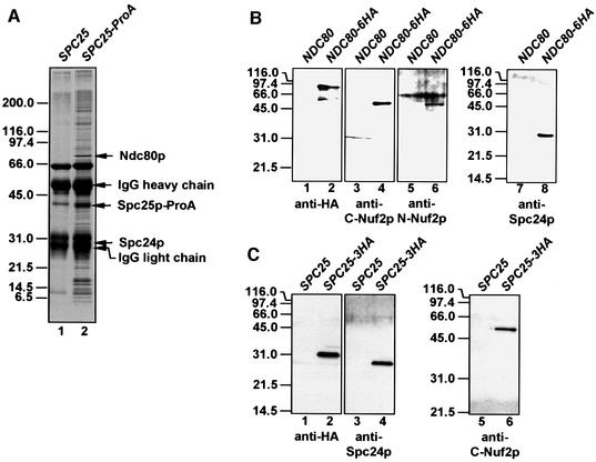
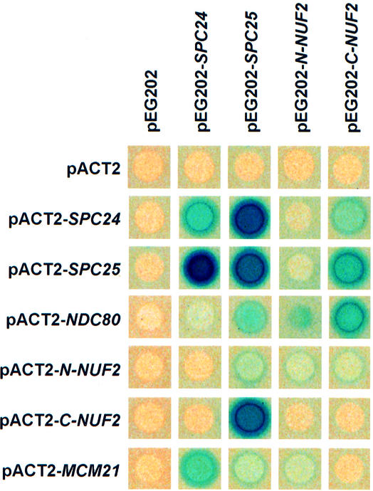
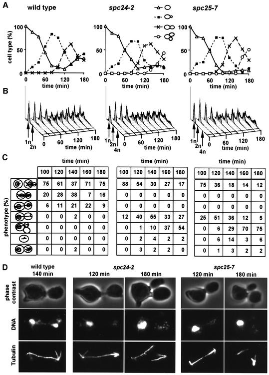
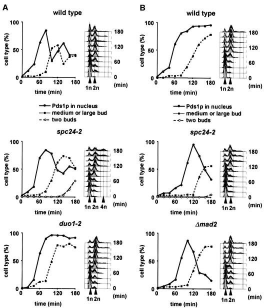
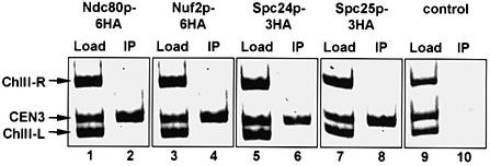
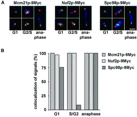
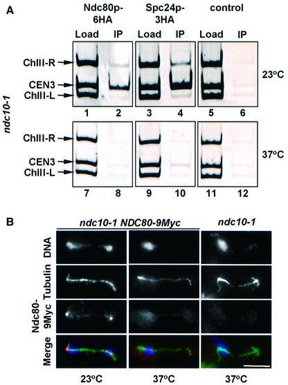
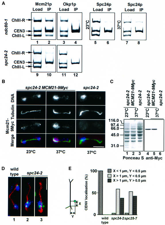
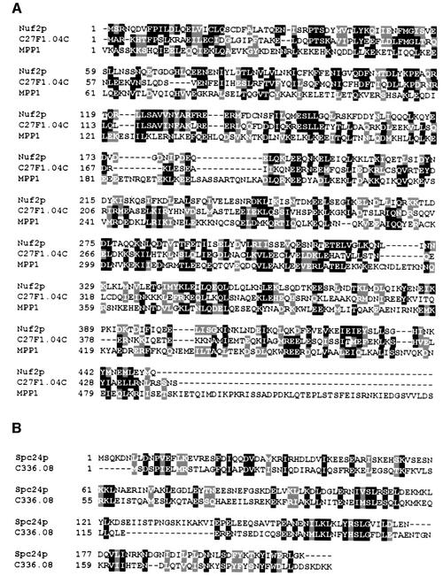
Here, we show that the budding yeast proteins Ndc80p, Nuf2p, Spc24p and Spc25p interact at the kinetochore. Consistently, Ndc80p, Nuf2p, Spc24p and Spc25p associate with centromere DNA in chromatin immunoprecipitation experiments, and SPC24 interacts genetically with MCM21 encoding a kinetochore component. Moreover, although conditional lethal spc24-2 and spc25-7 cells form a mitotic spindle, the kinetochores remain in the mother cell body and fail to segregate the chromosomes. Despite this defect in chromosome segregation, spc24-2 and spc25-7 cells do not arrest in metaphase in response to checkpoint control. Furthermore, spc24-2 cells showed a mitotic checkpoint defect when microtubules were depolymerized with nocodazole, indicating that Spc24p has a function in checkpoint control. Since Ndc80p, Nuf2p and Spc24p are conserved proteins, it is likely that similar complexes are part of the kinetochore in other organisms.


Figures

None
Fig. 1. Ndc80p, Nuf2p, Spc24p and Spc25p are present in common complexes. (A) Purification of Spc25p-associated proteins. Magnetic beads coated with rabbit IgGs were incubated with a yeast extract of wild-type (lane 1) or SPC25–ProA cells (lane 2). Bound proteins were eluted and separated by SDS–PAGE. Coomassie Blue-stained proteins were analysed by MALDI analysis. The major protein bands were identified as Ndc80p, Spc25p–ProA and Spc24p. The minor bands were contaminants like ribosomal proteins. (B) Co-immunoprecipitation of Ndc80p, Nuf2p and Spc24p. Extracts of NDC80 (lanes 1, 3, 5 and 7) or NDC80-6HA cells (lanes 2, 4, 6 and 8) were incubated with anti-HA beads. The precipitate was analysed by immunoblotting using anti-HA (lanes 1 and 2), anti-C-Nuf2p (lanes 3 and 4), anti-N-Nuf2p (lanes 5 and 6) and anti-Spc24p antibodies (lanes 7 and 8). (C) Co-immunoprecipitation of Nuf2p and Spc24p with Spc25p-6HA. The experimental design was as in (B), using SPC25 (lanes 1, 3 and 5) and SPC25-3HA cell extract (lanes 2, 4 and 6). The precipitates were tested with anti-HA (lanes 1 and 2), anti-Spc24p (lanes 3 and 4) and anti-C-Nuf2p antibodies (lanes 5 and 6).
None
Fig. 2. Ndc80p, Nuf2p, Mcm21p, Spc24p and Spc25p interact in the yeast two-hybrid system. Yeast strains containing the indicated yeast two-hybrid plasmids were overlayed with top agar containing 5-bromo-4-chloro-3-indolyl-β-d-galactoside (X-Gal) to measure β-galactosidase activity. Plates were incubated for 6 h at 30°C. Blue colour indicates interaction.
None
Fig. 3. spc24-2 and spc25-7 cells form an anaphase spindle but fail to segregate the chromosomes. Wild-type, spc24-2 and spc25-7 cells were synchronized with α-factor. The block was released and the cells were shifted to the restrictive temperature. Samples were withdrawn every 20 min and analysed for (A) budding index and (B) DNA content by flow cytometry. (C and D) Indirect immunofluorescence with anti-tubulin antibodies and 4′-6-diamidine-2-phenylindole (DAPI) staining. (C) is the quantification of (D) (n = 200). (C) In the cartoons at the left side of the panel, the grey circles within the cells symbolize the DAPI staining regions, and the lines the microtubules. Note that only the cell types after 100 min incubation at 37°C are shown.
None
Fig. 4. spc24-2 cells are checkpoint deficient. (A and B) α-factor synchronized wild-type, spc24-2, duo1-2 or Δmad2 cells containing PDS1-6HA were shifted to 37°C with (B) or without (A) 15 µg/ml nocodazole. Samples were withdrawn every 20 min and analysed for budding index, nuclear Pds1p by immunofluorescence, and DNA content by flow cytometry. Cells were also stained with anti-tubulin antibodies to ensure microtubule depolymerization when incubated with nocodazole.
None
Fig. 5. Ndc80p, Nuf2p, Spc24p and Spc25p are associated with the budding yeast kinetochore. NDC80-6HA (lanes 1 and 2), NUF2-6HA (lanes 3 and 4), SPC24-3HA (lanes 5 and 6) and SPC25-3HA cells (lanes 7 and 8), and wild-type cells without an HA tag (lanes 9 and 10) were subjected to ChIP. The immunoprecipitation was tested for the presence of CEN3 DNA and fragments left (ChIII-L) and right (ChIII-R) of CEN3 (IP: lanes 2, 4, 6, 8 and 10), as described previously (Ortiz et al., 1999). As a positive control for the PCR reactions, yeast lysates were tested without anti-HA precipitation (Load: lanes 1, 3, 5, 7 and 9).
None
Fig. 6. Nuf2p and Mcm21p are located close to the CEN5. (A) Localization of Mcm21p-9Myc, Nuf2p-9Myc and Spc98p-9Myc relative to CEN5 DNA. Synchronized cells with GFP-labeled CEN5 DNA carrying MCM21-9Myc, NUF2-9Myc or SPC98-9Myc were fixed and analysed by direct and indirect immunofluorescence for CEN5 DNA (green), Myc-tagged protein (red) and tubulin (blue). Note that the overlap between the green and red signals appears in yellow. Shown are cells in G1 phase (monopolar spindle), G2/S phase (short spindle) and anaphase (long spindle) of the cell cycle. Bar: 5 µm. (B) Quantification of (A). Cells (n = 100) in G1, G2/S phase or anaphase of the cell cycle were analysed. Signals were counted as ‘co-localized’ when the CEN5 signal (green) was <0.4 µm from the red anti-HA dot.
None
Fig. 7. Localization of Ndc80p and Spc24p in ndc10-1 cells. (A) Ndc80p and Spc24p are not associated with the kinetochore in ndc10-1 cells incubated at the restrictive temperature. Cells of ndc10-1 NDC80-6HA (lanes 1, 2, 7 and 8), ndc10-1 spc24-3HA (lanes 3, 4, 9 and 10) and ndc10-1 (control; lanes 5, 6, 11 and 12) grown at 23°C (lanes 1–6) were shifted to 37°C for 4 h (lanes 7–12). CEN3 association of Ndc80p-6HA (lanes 2 and 8) and Spc24p-3HA (lanes 4 and 10) was investigated by ChIP. ndc10-1 cells were employed as a control for the specificity of the immunoprecipitation (lanes 6 and 12) as well as binding to regions left (ChIII-L) and right (ChIII-R) of CEN3 DNA. Yeast lysates were also tested without precipitation (Load: lanes 1, 3, 5, 7, 9 and 11). (B) Cells of ndc10-1 NDC80-9Myc and ndc10-1 NDC80 were shifted from 23 to 37°C for 4 h. Fixed cells were analysed by indirect immunofluorescence using anti-tubulin and anti-Myc antibodies. DNA was stained with DAPI. The bottom panel shows the merged signals of the DNA (blue), tubulin (green) and Ndc80p-9Myc (red) stainings. Bar: 5 µm.
None
Fig. 8. Kinetochores are no longer clustered near SPBs in spc24-2 cells. (A) Mcm21p, Okp1p and Spc24p are associated with CEN3 DNA in ndc80-1 and spc24-2 cells. Cells of ndc80-1 (lanes 1–8) and spc24-2 (lanes 9–12) were tested for Mcm21p (lanes 1, 2, 9 and 10), Okp1p (lanes 3, 4, 11 and 12) and Spc24p (lanes 5–8) CEN3 DNA localization by ChIP at 23°C (lanes 5 and 6), or after 3 h at 37°C (lanes 1–4 and 7–12) using anti-Mcm21p (lanes 2 and 10), anti-Okp1p (lanes 4 and 12) or anti-Spc24p antibodies (lanes 6 and 8). Binding to regions left (ChIII-L) and right (ChIII-R) of CEN3 was tested as controls. Yeast lysates were assayed without precipitation (Load: lanes 1, 3, 5, 7, 9 and 11). (B) Localization of Mcm21p-9Myc in spc24-2 cells. Cells of spc24-2 MCM21-9Myc and spc24-2 were shifted from 23 to 37°C for 3 h. Fixed cells were analysed by indirect immunofluorescence using anti-Myc and anti-tubulin antibodies. DNA was stained with DAPI. The three signals were merged to compare localization. DAPI is shown in blue, the Mcm21p signal in red and microtubules in green. (C) Mcm21p levels remain constant. spc24-2 (lanes 3 and 6) and spc24-2 MCM21-9Myc cells (lanes 1, 2, 4 and 5) were grown at 23°C. One-half of the culture was incubated for 3 h at 37°C (lanes 2, 3, 5 and 6). Cell extracts were separated by SDS–PAGE and transferred to a nitrocellulose membrane. The membrane was stained with Ponceau S (lanes 1–3) to visualize proteins and then processed with anti-Myc antibodies (lanes 4–6). (D and E) Synchronized wild-type, spc24-2 and spc25-7 cells with GFP-labelled CEN5 DNA were shifted to 37°C. Cells were processed by anti-tubulin immunofluorescence and then analysed by confocal microscopy. (D) Large budded wild-type (panel 1) or spc24-2 cells (panels 2 and 3) containing an anaphase spindle. Tubulin is shown in red, CEN5 in green and DAPI in blue. (E) The position of CEN5 (green dot in cartoon) relative to the spindle pole (intersection of astral and nuclear microtubules) in the mother cell body of cells with an anaphase spindle was determined. Each CEN5 signal was counted separately. At least 100 cells were analysed. (B and D) Bars: 5 µm.
None
Fig. 9. Alignment of homologues of Nuf2p and Spc24p (program Clustal_X; Thompson et al., 1997). Homologous proteins were identified in a BLAST search (program WU-blastp; database, swall; matrix, blosum62; Altschul and Gish, 1996) using the protein sequence of Nuf2p and Spc24p. (A) The S.pombe homologue of Nuf2p, C27F1.04C [P(N) of 1.1 × 10⁻²⁷; high score of 318], is a hypothetical 51.9 kDa protein. The human MPP1 protein is homologue of Nuf2p [P(N) of 1.3 × 10⁻¹⁰; high score of 183]. Note that MPP1 is a protein of 225 kDa (Fritzler et al., 2000). (B) The S.pombe homologue of Spc24p, C336.08, is a hypothetical 23.0 kDa protein [P(N) of 2.1 × 10⁻¹⁰; high score of 148]. Identical amino acids are highlighted in black and similar amino acids in grey.

References

    1. Abrieu A., Kahana,J.A., Wood,K.W. and Cleveland,D.W. (2000) CENP-E as an essential component of the mitotic checkpoint in vitro. Cell, 102, 817–826. - PubMed
    1. Altschul S.F. and Gish,W. (1996) Local alignment statistics. Methods Enzymol., 266, 460–480. - PubMed
    1. Bardin A.J., Visintin,R. and Amon,A. (2000) A mechanism for coupling exit from mitosis to partitioning of the nucleus. Cell, 102, 21–31. - PubMed
    1. Chan G.K., Jablonski,S.A., Sudakin,V., Hittle,J.C. and Yen,T.J. (1999) Human BUBR1 is a mitotic checkpoint kinase that monitors CENP-E functions at kinetochores and binds the cyclosome/APC. J. Cell Biol., 146, 941–954. - PMC - PubMed
    1. Chen R.H., Waters,J.C., Salmon,E.D. and Murray,A.W. (1996) Association of spindle assembly checkpoint component XMAD2 with unattached kinetochores. Science, 274, 242–246. - PubMed

Publication types

MeSH terms