. 2001 Feb 15;20(4):792-801.
doi: 10.1093/emboj/20.4.792.

Securin degradation is mediated by fzy and fzr, and is required for complete chromatid separation but not for cytokinesis

Affiliations

Securin degradation is mediated by fzy and fzr, and is required for complete chromatid separation but not for cytokinesis

A Zur et al. EMBO J. .

Abstract

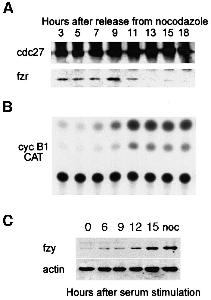
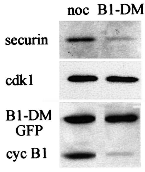
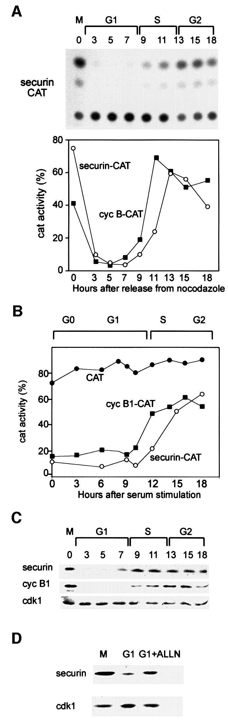
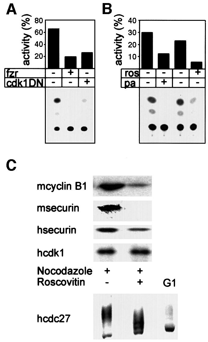
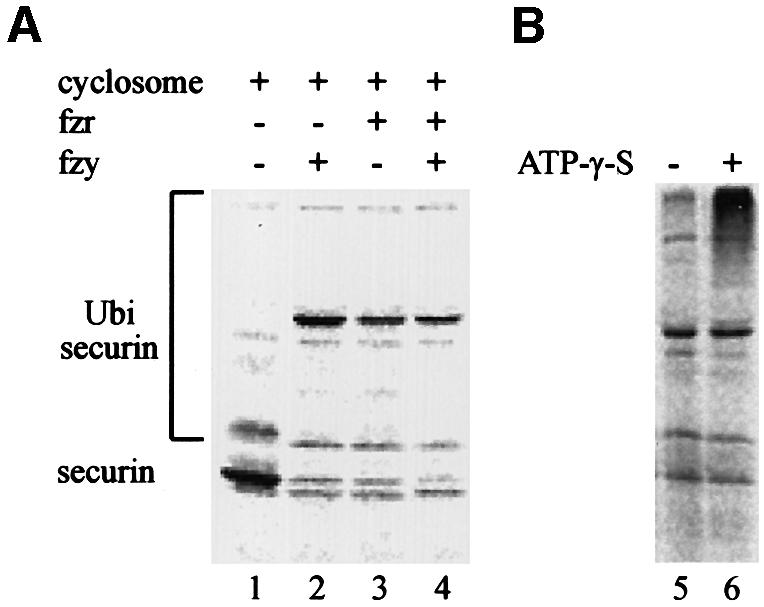
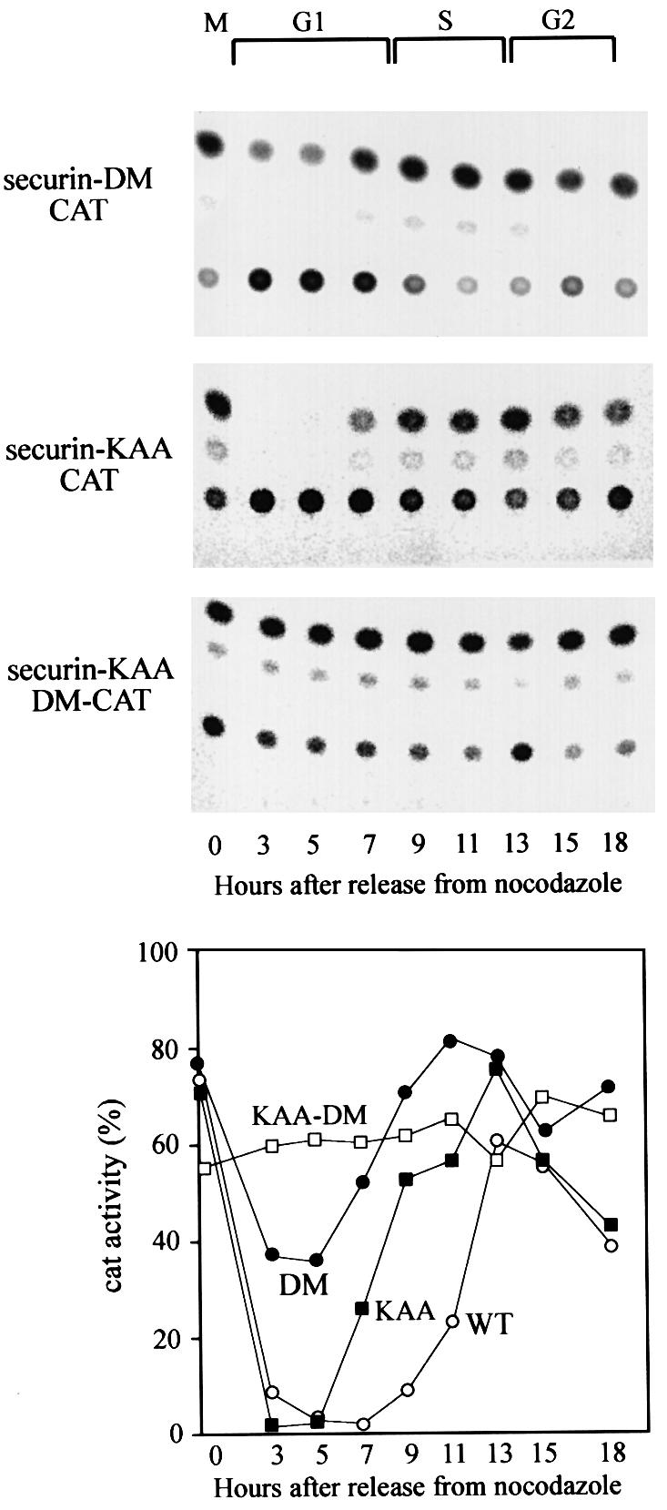
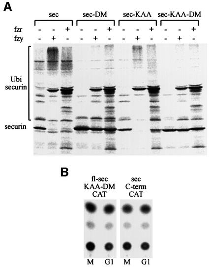
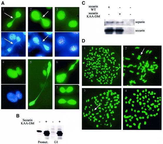
We have studied the ubiquitination and degradation patterns of the human securin/PTTG protein. We show that, in contrast to budding yeast pds1, securin degradation is catalyzed by both fzy (fizzy/cdc20) and fzr (fizzy-related/cdh1/hct1). Both fzy and fzr also induce the APC/C to ubiquitinate securin in vitro. Securin degradation is mediated by an RXXL destruction box and a KEN box, and is inhibited only when both sequences are mutated. Interestingly, the non-degradable securin mutant is also partially ubiquitinated by fzy and fzr in vitro. Expressing the non-degradable securin mutant in cells frequently resulted in incomplete chromatid separation and gave rise to daughter cells connected by a thin chromatin fiber, presumably of chromosomes that failed to split completely. Strikingly, the mutant securin did not prevent the majority of sister chromatids from separating completely, nor did it prevent mitotic cyclin degradation and cytokinesis. This phenotype, reminiscent of the fission yeast cut (cells untimely torn) phenotype, is reported here for the first time in mammals.


Figures

None
Fig. 1. Fzr binding to the APC/C is associated with its G1-specific activity. (A) NIH 3T3 cells stably expressing cyclin B1–CAT were synchronized at prometaphase with nocodazole and subsequent shake-off of the rounded cells. They were washed and released into fresh medium and harvested at the indicated time points. Cells synchronized by this method pass the G1–S transition at ∼7–9 h. Cell extracts were immunoprecipitated with anti-cdc27 beads. The immunoprecipitates were analyzed by immunoblotting with fzr and cdc27 antibodies. (B) The cells were analyzed further for CAT activity, an indication of the stability of the cyclin B1–CAT and thus of APC/C activity or inactivity. (C) Serum-starved, G0-arrested fibroblasts were stimulated with serum to re-enter the cell cycle and were harvested at the indicated time points. They were immunoblotted with fzy antibodies and with α-actin antibodies which served as a loading control. Entry into S phase takes place at 12–15 h after stimulation. An extract of nocodazole-arrested cells, at a time when fzy levels peak in the cell, was used as a positive control.
None
Fig. 2. APC/Cfzy-mediated degradation of securin. Cells were transfected with non-degradable GFP–cyclin B1 and either arrested in prometaphase with nocodazole or allowed to proceed and arrest in telophase. Cells were obtained by mitotic shake-off, and securin and cyclin B1 levels were analyzed by immunoblotting. Cdk1, which is stable in G1, served as a loading control.
None
Fig. 3. Securin and cyclin B1 have a similar degradation pattern in G1. (A) Cells were stably transfected with the securin–CAT expression vector which constitutively transcribes a reporter fusion protein between the N-terminal 87 residues of human securin and a bacterial CAT protein. Cells were synchronized as described and assayed for CAT activity. The CAT activity was calculated as the ratio of the amount of diacetylated [14C]chloramphenicol to that of total [14C]chloramphenicol (diacetylated + non-acetylated) as quantified by a phosphoimager. The changes in the activities of securin–CAT (open circles) and cyclin B1–CAT (filled squares) (shown in Figure 1) were plotted as a function of the cell cycle phases when released from a nocodazole block. (B) Cells stably expressing CAT (filled circles), cyclin B1–CAT (filled squares) and securin–CAT (open circles) were arrested by serum deprivation in G0 and released by serum stimulation. Cells were harvested and analyzed for CAT activity at the indicated time points. (C) Cells were synchronized at prometaphase by nocodazole arrest and mitotic shake-off, released into fresh medium and harvested at the indicated time points for immunoblotting with securin, cyclin B1 and cdk1 antibodies. (D) Prometaphase-arrested cells were washed and released into fresh medium for 2 h, and then either harvested or grown for an additional 2 h with or without ALLN. Cell extracts were immunoblotted with securin or cdk1 antibodies. Cdk1 is constant in G1 and served as a loading control.
None
Fig. 4. Fzr overexpression and cdk1 inactivation lead to securin degradation in prometaphase. (A) Cells were co-transfected with securin–CAT with a fzr expression vector, a cdk1DN expression vector or an empty vector. They were arrested with nocodazole, harvested by shake-off and analyzed for CAT activity. Both fzr overexpression and cdk1 inhibition strongly activated the APC/C-specific degradation of securin–CAT in prometaphase. (B) Cells expressing securin–CAT were arrested in prometaphase by nocodazole and subsequently treated for 3 h with the cdk inhibitors roscovitin and purvalanol A, in the presence of nocodazole. They were then assayed for CAT activity. (C) Mouse (m) NIH 3T3 fibroblasts or human (h) HeLa cells were treated as in (B) and immunoblotted with securin, cyclin B1, cdk1 and cdc27 antibodies. The last antibody was used to show that roscovitin indeed leads to cdk1 inhibition in vivo as seen by the decrease in the phosphorylation of cdc27, a known cdk1 phosphorylation substrate.
None
Fig. 5. Both fzy and fzr can activate the APC/C to ubiquitinate securin. (A) An in vitro ubiquitination assay was performed on 35S-labeled securin expressed in reticulocyte lysate (lane 1) with a partially purified APC/C from mitotic HeLa cells in the presence of bacterially expressed E1, E2-C, an energy-regenerating system and ubiquitin aldehyde, which inhibits de-ubiquitination. This reaction was supplemented with either fzy (lane 2), fzr (lane 3) or both (lane 4). (B) Some of the ubiquitinated securin must have been degraded by the proteasome in the reticulocyte lysate. We therefore repeated the ubiquitination reaction in the presence (lane 6) or absence (lane 5) of ATP-γ-S, which enables ubiquitination but not proteasome-specific degradation.
None
Fig. 6. Securin has two conserved sequences that signal its APC/C-specific degradation. Cells stably expressing securin-DM–CAT (filled circles), which carries a mutated d-box, securin-KAA–CAT (filled squares) with a mutated KEN box and securin-KAA-DM–CAT (open squares) with a double mutation were generated. These cells were released from a nocodazole block, harvested at the indicated time points and the CAT assays are shown in the top panels. The quantified CAT activity of the above mutants, as well as of wild-type securin–CAT (open circles), shown in Figure 3, is plotted in the bottom panel.
None
Fig. 7. Both degradable and non-degradable securin mutants are ubiquitinated by the APC/C. (A) Full-length securin–CAT, securin-DM–CAT, securin-KAA–CAT and securin-KAA-DM–CAT were expressed in reticulocyte lysate and tested for ubiquitination as described in the legend to Figure 5, in the presence of ATP-γ-S. (B) Plasmids expressing either full-length non-degradable securin–CAT (fl-sec-KAA-DM–CAT) or the C-terminal half of securin fused to CAT (sec-C-term–CAT) were transfected into cells. Cells were arrested in prometaphase and were either harvested immediately for CAT assay or first released into G1 for 3 h.
None
Fig. 8. Expression of indestructible securin interferes with chromatid separation. (A) NIH 3T3 cells were transfected with vectors expressing non-degradable (panels 1, 2, 4, 5 and 6) and wild-type (panel 3) securin together with a GFP–histone H2A expression vector (green panels). Cells were stained further in vivo with Hoechst 33258 (blue panels). About 1/3 of the cells expressing the mutant securin failed to separate all their chromatids prior to cytokinesis and remained connected by a thin chromatin string (arrow), or even failed to complete nuclear division altogether (panel 4). The same phenotype was observed in some HeLa cells expressing non-degradable securin (panel 7). (B) Cells transfected with an empty vector or with non-degradable securin were arrested in prometaphase and either harvested immediately or released first into G1 for 3 h. Cell extracts subsequently were immunoblotted with securin antibodies. (C) Cells were transfected with an expression vector for myc-tagged wild-type and non-degradable securin. Extracts prepared from transfected and untransfected cells were immunoprecipitated with myc tag (9E10) antibodies and immunoblotted with separin and securin antibodies. (D) Cells were co-transfected with expression vectors for securin (wild-type or non-degradable), non-degradable cyclin B1 and GFP–H2A. Cells were arrested in mitosis and were harvested by shake-off. Chromosome spreads were prepared as described in Materials and methods. Cells transfected with wild-type, and the majority of those transfected with non-degradable securin, completely dissociated chromatids (panel 1). However, many of those transfected with non-degradable securin still had a few (panel 2; arrow shows a separated chromosome), most (panel 3; arrow shows an unseparated chromosome) or all (panel 4) chromatids paired.

References

    1. Amon A., Irniger,S. and Nasmyth,K. (1994) Closing the cell cycle circle in yeast: G2 cyclin proteolysis initiated at mitosis persists until the activation of G1 cyclins in the next cycle. Cell, 77, 1037–1050. - PubMed
    1. Antonio C., Ferby,I., Wilhelm,H., Jones,M., Karsenti,E., Nebreda,A. and Vernos,I. (2000) Xkid, a chromokinesin required for chromosome alignment on the metaphase plate. Cell, 102, 425–435. - PubMed
    1. Ausubel F.M., Brent,R., Kingston,R.E., Moore,D.D., Seidman,J.G., Smith,J.A. and Struhl,K. (1994) Current Protocols in Molecular Biology. John Wiley & Sons Inc., New York, NY.
    1. Brandeis M. and Hunt,T. (1996) The proteolysis of mitotic cyclins in mammalian cells persists from the end of mitosis until the onset of S phase. EMBO J., 15, 5280–5289. - PMC - PubMed
    1. Ciosk R., Zachariae,W., Michaelis,C., Shevchenko,A., Mann,M. and Nasmyth,K. (1998) An ESP1/PDS1 complex regulates loss of sister chromatid cohesion at the metaphase to anaphase transition in yeast. Cell, 93, 1067–1076. - PubMed

Publication types