. 2001 Mar;69(3):1581-6.
doi: 10.1128/IAI.69.3.1581-1586.2001.

Staphylococcus aureus and Salmonella enterica serovar Dublin induce tumor necrosis factor-related apoptosis-inducing ligand expression by normal mouse and human osteoblasts

Affiliations

Staphylococcus aureus and Salmonella enterica serovar Dublin induce tumor necrosis factor-related apoptosis-inducing ligand expression by normal mouse and human osteoblasts

E H Alexander et al. Infect Immun. 2001 Mar.

Abstract

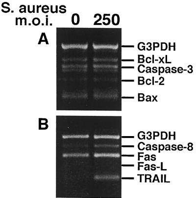
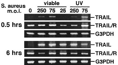
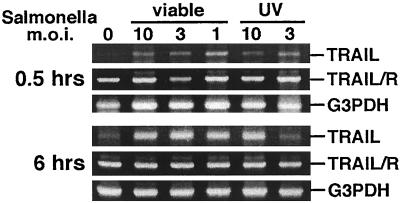
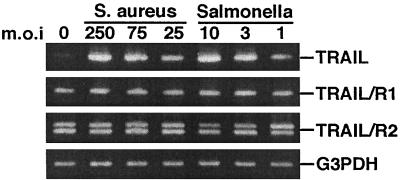
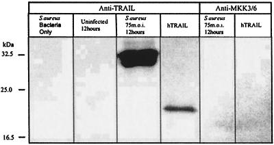
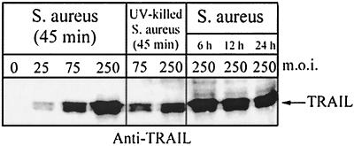
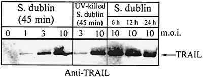
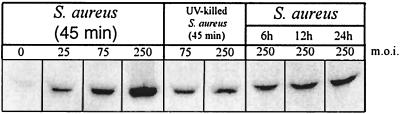
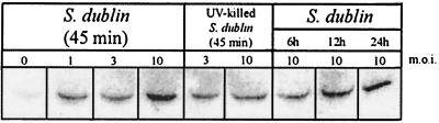
Staphylococcus aureus and Salmonella enterica serovar Dublin invade osteoblasts and are causative agents of human bone disease. In the present study, we examined the ability of S. aureus and Salmonella serovar Dublin to induce the production of tumor necrosis factor-related apoptosis-inducing ligand (TRAIL) by normal osteoblasts. Normal mouse and human osteoblasts were cocultured with S. aureus or Salmonella serovar Dublin at different multiplicities of infection. Following initial incubation and examination of TRAIL expression, extracellular bacteria were killed by the addition of media containing the antibiotic gentamicin. Lysates and conditioned media from osteoblast cultures were then collected at various times following invasion and analyzed. The results demonstrated that S. aureus and Salmonella serovar Dublin are potent inducers of TRAIL expression by osteoblasts. Mouse and human TRAIL mRNA expression was induced by bacterial infection and demonstrated a dose-dependent response. Analysis of kinetics suggested that TRAIL mRNA was induced within 30 min after exposure to bacteria and that its level of expression remained relatively constant over the time period examined. mRNA molecules encoding TRAIL receptors were constitutively expressed by osteoblasts. Furthermore, TRAIL protein was detected as early as 45 min and up to 24 h following infection. The quantity of TRAIL protein produced also increased in a dose-dependent manner. Collectively, these findings suggest a mechanism whereby bacterial pathogens mediate bone destruction via osteoblast apoptosis.


Figures

FIG. 1
FIG. 1
RT-PCR analysis of apoptosis-related genes in normal human osteoblasts infected with S. aureus at an MOI of 250:1 (S. aureus organisms to osteoblasts). PCR amplification was performed as recommended by BioSource International. Gene expression was monitored at 6 h following the addition of viable bacteria. Bcl-xL, B-cell lymphoma gene × Long; Bcl-2, B-cell lymphoma 2; Bax, Bcl-2-associated × protein; Fas, CD95; Fas-L, Fas ligand.
FIG. 2
FIG. 2
RT-RCR analysis of TRAIL and TRAIL receptor (TRAIL/R) expression in normal mouse osteoblasts infected with S. aureus or UV-killed S. aureus at different MOIs (25:1, 75:1, or 250:1 [S. aureus organisms to osteoblasts]). PCR amplification of G3PDH was performed to ensure equal input of RNA and similar efficiencies of RT. Gene expression was monitored at 0.5 and 6 h following the addition of viable or UV-killed bacteria.
FIG. 3
FIG. 3
RT-RCR analysis of TRAIL and TRAIL receptor (TRAIL/R) expression in normal mouse osteoblasts infected with Salmonella serovar Dublin or UV-killed Salmonella serovar Dublin at different MOIs (10:1, 3:1, or 1:1 [Salmonella serovar Dublin organisms to osteoblasts]). PCR amplification of G3PDH was performed to ensure equal input of RNA and similar efficiencies of RT. Gene expression was monitored at 0.5 and 6 h following the addition of viable or UV-killed bacteria.
FIG. 4
FIG. 4
RT-RCR analysis of TRAIL, TRAIL R1, and TRAIL R2 expression in normal human osteoblasts infected with S. aureus or Salmonella serovar Dublin at different MOIs (25:1, 75:1, or 250:1 [S. aureus organisms to osteoblasts]; 1:1, 3:1, or 10:1 [Salmonella serovar Dublin organisms to osteoblasts]). PCR amplification of G3PDH was performed to ensure equal input of RNA and similar efficiencies of RT. Gene expression was monitored at 6 h following the addition of viable bacteria.
FIG. 5
FIG. 5
Western immunoblot analysis of cell-associated TRAIL expression by normal human osteoblasts infected with S. aureus. Lanes contained S. aureus devoid of osteoblasts; uninfected osteoblasts 12 h after mock infection; osteoblasts infected with S. aureus at an MOI of 75:1, 12 h after the addition of bacteria; and recombinant human TRAIL (hTRAIL). The first four lanes were probed with polyclonal anti-TRAIL antibody, and the fifth and sixth lanes were probed with polyclonal anti-phosphorylated MKK3 or MKK6 antibody, prior to reaction with the secondary antibody. Two micrograms of total protein was loaded for the first three lanes and the fifth lane; 1 μg of recombinant hTRAIL was loaded for the fourth and sixth lanes.
FIG. 6
FIG. 6
Western immunoblot analysis of cell-associated TRAIL expression by normal human osteoblasts infected with S. aureus or UV-killed S. aureus at different MOIs (25:1, 75:1, or 250:1 [S. aureus organisms to osteoblasts]). TRAIL expression was monitored at 45 min following the addition of viable or UV-killed bacteria and at 6, 12, and 24 h following infection with viable S. aureus cells. Two micrograms of total protein was loaded for each lane.
FIG. 7
FIG. 7
Western immunoblot analysis of cell-associated TRAIL expression by normal human osteoblasts infected with Salmonella serovar Dublin or UV-killed Salmonella serovar Dublin at different MOIs (1:1, 3:1, or 10:1 [Salmonella serovar Dublin organisms to osteoblasts]). TRAIL expression was monitored at 45 min following the addition of viable or UV-killed bacteria and at 6, 12, and 24 h following infection with viable Salmonella serovar Dublin cells. Two micrograms of total protein was loaded for each lane.
FIG. 8
FIG. 8
Western immunoblot analysis of TRAIL secretion by normal human osteoblasts infected with S. aureus or UV-killed S. aureus at different MOIs. Lanes, from left to right, contained protein reactive with anti-TRAIL antibody in culture supernatants 45 min following infection with S. aureus at an MOI of 0, 25:1, 75:1, or 250:1; protein reactive with anti-TRAIL antibody in culture supernatants 45 min following the addition of UV-killed S. aureus at an MOI of 75:1 or 250:1; and protein reactive with anti-TRAIL antibody in culture supernatants at 6, 12, and 24 h following infection with S. aureus at an MOI of 250:1. Ten micrograms of total protein from serum-containing medium was loaded for each lane.
FIG. 9
FIG. 9
Western immunoblot analysis of TRAIL secretion by normal human osteoblasts infected with Salmonella serovar Dublin or UV-killed Salmonella serovar Dublin at different MOIs. Lanes, from left to right, contained protein reactive with anti-TRAIL antibody in culture supernatants 45 min following infection with Salmonella serovar Dublin at an MOI of 0, 1:1, 3:1, or 10:1; protein reactive with anti-TRAIL antibody in culture supernatants 45 min following the addition of UV-killed Salmonella serovar Dublin at an MOI of 3:1 or 10:1; and protein reactive with anti-TRAIL antibody in culture supernatants at 6, 12, and 24 h following infection with Salmonella serovar Dublin at an MOI of 10:1. Ten micrograms of total protein from serum-containing medium was loaded for each lane.

References

    1. Almeida R A, Matthews K R, Cifrian E, Guidry A J, Oliver S P. Staphylococcus aureus invasion of bovine mammary epithelial cells. J Dairy Sci. 1996;79:1021–1026. - PubMed
    1. Bayles K W, Wesson C A, Liou L E, Fox L K, Bohach G A, Trumble W R. Intracellular Staphylococcus aureus escapes the endosome and induces apoptosis in epithelial cells. Infect Immun. 1998;66:336–342. - PMC - PubMed
    1. Beekhuizen H, Van De Gevel J S, Olsson B, Van Benten I J, Van Furth R. Infection of human vascular endothelial cells with Staphylococcus aureus induces hyperadhesiveness for human monocytes and granulocytes. J Immunol. 1997;158:774–782. - PubMed
    1. Bost K L, Clements J D. In vivo induction of interleukin-12 mRNA expression after oral immunization with Salmonella dublin or the B subunit of Escherichia coli heat-labile enterotoxin. Infect Immun. 1995;63:1076–1083. - PMC - PubMed
    1. Bost K L, Mason M J. Thapsigargin and cyclopiazonic acid initiate rapid and dramatic increases in IL-6 mRNA expression and IL-6 secretion in murine peritoneal macrophages. J Immunol. 1995;155:285–296. - PubMed

Publication types

MeSH terms

Substances

LinkOut - more resources