. 2001 Feb 15;531(Pt 1):131-40.
doi: 10.1111/j.1469-7793.2001.0131j.x.

Intracellular pathways regulating ciliary beating of rat brain ependymal cells

Affiliations

Intracellular pathways regulating ciliary beating of rat brain ependymal cells

T Nguyen et al. J Physiol. .

Abstract

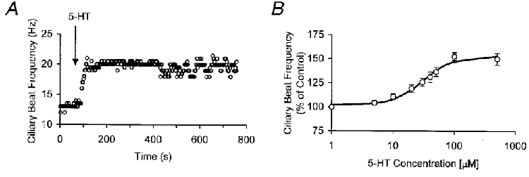
1. The mammalian brain ventricles are lined with ciliated ependymal cells. As yet little is known about the mechanisms by which neurotransmitters regulate cilia beat frequency (CBF). 2. Application of 5-HT to ependymal cells in cultured rat brainstem slices caused CBF to increase. 5-HT had an EC50 of 30 microM and at 100 microM attained a near-maximal CBF increase of 52.7 +/- 4.1 % (mean +/- s.d.) (n = 8). 3. Bathing slices in Ca2+-free solution markedly reduced the 5-HT-mediated increase in CBF. Fluorescence measurements revealed that 5-HT caused a marked transient elevation in cytosolic Ca2+ ([Ca2+]c) that then slowly decreased to a plateau level. Analysis showed that the [Ca2+]c transient was due to release of Ca2+ from inositol 1,4,5-trisphosphate (IP3)-sensitive stores; the plateau was probably due to extracellular Ca2+ influx through Ca2+ release-activated Ca2+ (CRAC) channels. 4. Application of ATP caused a sustained decrease in CBF. ATP had an EC50 of about 50 microM and 100 microM ATP resulted in a maximal 57.5 +/- 6.5 % (n = 12) decrease in CBF. The ATP-induced decrease in CBF was unaffected by lowering extracellular [Ca2+], and no changes in [Ca2+]c were observed. Exposure of ependymal cells to forskolin caused a decrease in CBF. Ciliated ependymal cells loaded with caged cAMP exhibited a 54.3 +/- 7.5 % (n = 9) decrease in CBF following uncaging. These results suggest that ATP reduces CBF by a Ca2+-independent cAMP-mediated pathway. 5. Application of 5-HT and adenosine-5'-O-3-thiotriphosphate (ATP-gamma-S) to acutely isolated ciliated ependymal cells resulted in CBF responses similar to those of ependymal cells in cultured slices suggesting that these neurotransmitters act directly on these cells. 6. The opposite response of ciliated ependymal cells to 5-HT and ATP provides a novel mechanism for their active involvement in central nervous system signalling.


Figures

Figure 1
Figure 1. 5-HT immunolabelling in transverse rat brainstem slices
A, tryptophan hydroxylase-positive processes are present at the ependymal cell layer. Arrow points to the basal side of the ependymal cell layer where there are both 5-HT-containing nerve fibres and varicosities. Tissue section is from the medulla rostral to the obex where the 4th ventricle is clearly evident. This picture is derived from successive z-series images of the tissue section. B, tryptophan hydroxylase-positive neurons of the nucleus raphe obscurus are present in the same tissue section as in A. This picture is derived from single z-image of the tissue section. Calibration bars: 50 μm in A and 100 μm in B.
Figure 2
Figure 2. Effect of 5-HT on ciliary beating of rat ependymal cells
A, CBF response of an ependymal cell to 5-HT (50 μM) in normal Hanks’ solution. B, dose-response of CBF to varying 5-HT concentrations (n= 8). CBF increases with increasing 5-HT concentration with an EC50 of 30 μM and a near-maximal response at 100 μM. Data points are means ±s.d. and were fitted to a Hill equation.
Figure 3
Figure 3. Role of Ca2+ in the 5-HT-mediated increase of ependymal cell CBF
A, CBF response of an ependymal cell to 5-HT (50 μM) in EGTA-buffered Ca2+-free Hanks’ solution. The mean increase in CBF was markedly reduced to 5.6 ± 2.3 % in Ca2+-free medium (n= 12). B, effect of 5-HT (50 μM) in Hanks’ solution on [Ca2+]c. C, effect of 5-HT (50 μM) in EGTA-buffered Ca2+-free Hanks’ solution on [Ca2+]c.
Figure 4
Figure 4. Electrical properties of ependymal cells
A, photomicrograph of a Alexa-dye-filled ependymal cell that was recorded from in voltage-clamp mode. Lightly stained cilia are seen (*) extending into the 4th ventricle. Arrow points to a short basal process of the filled cell. Calibration bar is 40 μm. B, the steady-state current-voltage relationship is linear. C, whole-cell current recordings from two different holding potentials shows an absence of any inward currents. Membrane current responses to 10 mV incrementing steps from a −70 mV holding potential (left-hand panel) and from a −80 mV potential (right-hand panel). All data shown are from the same ependymal cell.
Figure 5
Figure 5. Xestospongin blockade of IP3 receptors abolishes the effect of 5-HT on CBF and [Ca2+]c
A, effect of xestospongin, a membrane-permeable IP3 receptor antagonist, on the 5-HT-mediated effect on CBF. Xestospongin (20 μM) completely abolished the effect of 5-HT (50 μM) on CBF. B, xestospongin (20 μM) completely abolished the effect of 5-HT on [Ca2+]c.
Figure 6
Figure 6. Effect of ATP on ciliary beating and [Ca2+]c of ependymal cells
A, CBF response of an ependymal cell to ATP (100 μM) in normal Hanks’ solution. B, dose-response of CBF to varying concentrations of exogenously applied ATP (n= 12). CBF decreases with ATP concentration, with an EC50 of approximately 50 μM, and attains a maximal decrease at a concentration of 100 μM. Error bars are s.d.s. C, effect on CBF of ATP (100 μM) and ATP-γ-S (100 μM), a non-hydrolysable ATP analogue, in the presence of 0.1 units ATPase (n= 6). In the presence of ATPase, ATP does not cause a decrease in CBF; in contrast, application of ATP-γ-S results in a decrease in CBF. D, measurement of ependymal cell [Ca2+]c exposed to ATP (100 μM) in Hanks’ solution. No changes in [Ca2+]c were observed with ATP application.
Figure 7
Figure 7. Effect of uncaging of cAMP on ciliary beating of ependymal cells
Effect of uncaging cAMP with a 240-270 nm light flash on CBF in an ependymal cell loaded with 50 μM caged cAMP. Releasing cAMP results in a 54 ± 7.5 % (n= 9) decrease in CBF.
Figure 8
Figure 8. Effect of 5-HT and ATP-γ-S on ciliary beating of isolated rat ependymal cells
A, application of 5-HT (50 μM) to an isolated ependymal cell resulted in an increase in CBF. B, application of ATP-γ-S (100 μM), a non-hydrolysable ATP analogue, caused a marked decreased in CBF.
Figure 9
Figure 9. A model of the signalling pathways involved in the effect of 5-HT and ATP on ciliary beating of ependymal cells
Exposure to 5-HT results in the intracellular production of IP3, which releases Ca2+ from intracellular Ca2+ stores. The released Ca2+ mediates the opening of CRAC channels that results in the influx of Ca2+ from the extracellular environment. The increase in [Ca2+]c ultimately increases CBF by an as yet unidentified mechanism. In contrast, ATP activates the production of intracellular cAMP, which inhibits CBF by a still to be determined mechanism.

References

    1. Afzelius BA, Mossberg B. Immotile-cilia syndrome (primary ciliary dyskinesia), including Kartagener syndrome. In: Scriver CR, Beaudet AL, Sly WS, Valle D, editors. The Metabolic and Molecular Bases of Inherited Disease. New York: McGraw-Hill; 1995. pp. 3943–3954.
    1. Ben-Shimol Y, Dinstein I, Meisels A, Priel Z. Ciliary motion features from digitized video photography. Journal of Computer-Assisted Microscopy. 1991;3:103–116.
    1. Böhm SK, Khitin LM, Grady EF, Aponte G, Payan DG, Bunnett NW. Mechanisms of desensitization and resensitization of proteinase-activated receptor-2. Journal of Biological Chemistry. 1996;271:22003–22016. - PubMed
    1. Cathcart RS, Worthington WC. Ciliary movement in the rat cerebral ventricles: clearing action and directions of currents. Journal of Neuropathology and Experimental Neurology. 1964;23:609–618. - PubMed
    1. Chan-Paly V. Serotonin axons in the supra- and subependymal plexuses and in the leptomeninges; their roles in local alterations of cerebrospinal fluid and vasomotor activity. Brain Research. 1976;102:103–130. - PubMed

Publication types

LinkOut - more resources