. 2001 Feb 15;531(Pt 1):265-75.
doi: 10.1111/j.1469-7793.2001.0265j.x.

Voluntary contraction impairs the refractory period of transmission in healthy human axons

Affiliations

Voluntary contraction impairs the refractory period of transmission in healthy human axons

S Kuwabara et al. J Physiol. .

Abstract

1. Voluntary contraction of a muscle causes substantial hyperpolarization of the active motor axons due to activation of the electrogenic Na+-K+ pump. The present study was undertaken to determine whether voluntary effort produces a significant impairment in impulse transmission in normal axons and whether mechanisms other than membrane hyperpolarization contribute to the changes in axonal excitability. 2. The compound muscle action potential (CMAP) was recorded after median nerve stimulation at the wrist using sub- and supramaximal stimuli, delivered singly and in pairs at conditioning-test intervals of 2-15 ms. Axonal excitability parameters (threshold, refractoriness, supernormality, and strength-duration time constant (tauSD)) were measured using threshold tracking. Impulse transmission was assessed using supramaximal stimuli. 3. Maximal voluntary contractions of the abductor pollicis brevis for 1 min produced a substantial increase in threshold, an increase in supernormality and a decrease in tauSD, all of which lasted approximately 10 min and indicate axonal hyperpolarization. However, immediately after the contraction there was an unexpected increase in refractoriness. The post-contraction increase in refractoriness could not be mimicked by an imposed ramp of hyperpolarization that produced changes in the other indices to an extent that was similar to voluntary contraction. 4. The contraction had relatively little effect on the size of the unconditioned maximal CMAP. However, there was failure of transmission of supramaximal conditioned volleys when the conditioning-test interval was short. 5. The relationships between axonal excitability and supernormality and tauSD following voluntary contraction differed significantly from those recorded during the hyperpolarization produced by DC current. It is argued that these differences probably result from extra-axonal K+ accumulation with the voluntary contraction but not with the DC polarization. I6. It is concluded that, following maximal voluntary contraction of a normal muscle, the refractory period of transmission is impaired distal to the stimulus site sufficient to cause transmission failure of the second of a pair of closely spaced impulses. The site of transmission failure is likely to be the terminal axon, presumably at branch points, possibly in the unmyelinated pre-terminal segment.


Figures

Figure 4
Figure 4. Changes in impulse transmission produced by maximal voluntary contraction for 1 min
A, changes in amplitude of unconditioned compound muscle action potential (CMAP) recorded from the abductor pollicis brevis in a single subject. To prove that stimulus intensity remained supramaximal after contraction, a second stimulus (stimulus 2) was introduced after the contraction. Stimulus 2 was set 20 % stronger than stimulus 1. B, amplitudes of conditioned CMAPs produced by supramaximal stimuli at conditioning–test intervals of 2–15 ms. C, unconditioned and conditioned CMAPs to supramaximal stimuli before maximal voluntary contraction. Traces are, from top to bottom, maximal CMAP to an unconditioned supramaximal stimulus, CMAP to an even stronger supramaximal stimulus, responses to supramaximal conditioning and test stimuli (interval 2 ms), the CMAP to the conditioned stimulus (determined by subtracting the unconditioned CMAP from the response to the conditioning and test stimulus pair). D, post-contraction changes in the conditioned CMAP (30, 60 and 90 s after the end of the contraction).
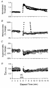
Figure 1
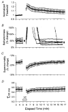
Figure 1. Changes in excitability produced by a 1 min maximal voluntary contraction (•) and a 10 % continuous hyperpolarizing current (○) in a single subject
A, threshold changes measured using test stimuli of 1.0 ms duration. B, refractoriness measured using a 0.1 ms test stimulus delivered 2 ms after a supramaximal conditioning stimulus. Note that following voluntary effort (VC in D, applies also to A-C) there was an abrupt increase in the conditioned threshold for 3 min, superimposed on a long-lasting decrease. The dashed line in B indicates the pre-contraction value. C, supernormality measured during the supernormal period using a 0.1 ms test stimulus delivered 7 ms after a supramaximal stimulus. In B and C, the change is expressed as a percentage of the unconditioned threshold. DSD calculated from the threshold changes using test stimuli of 0.1 and 1.0 ms duration.
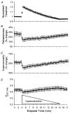
Figure 3
Figure 3. Changes in excitability indices produced by continuous hyperpolarizing currents
Figure layout as for Fig. 1. Means ±s.e.m. for 6 subjects. The intensity of artificial hyperpolarization was maximal (10 % of current required to produce a CMAP that was 50 % of maximum) at 5 min and gradually decreased over 10 min. Data have been averaged over consecutive 15 s intervals.
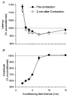
Figure 5
Figure 5. Latency-based recovery cycle of axonal excitability
A, latency of the CMAP, measured to half-peak, normalized to the latency of unconditioned response. Pre-contraction data were obtained over 2 min before the contraction, and post-contraction data from 2-4 min after the end of the contraction. B, amplitude of the conditioned CMAP to supramaximal stimuli. Note the recovery of latency is much quicker than that of amplitude. All data points represent means ±s.e.m. for 9 subjects. (The symbols are often larger than the error bars.)
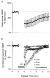
Figure 6
Figure 6. Changes in excitability (A) and CMAP amplitude (B) produced by maximal voluntary contraction for 1 min
Means ±s.e.m. for 9 subjects. A, amplitude of CMAP elicited by a constant submaximal stimulus which initially produced a CMAP 70 % of maximal. B, amplitudes of the conditioned CMAP produced by supramaximal test stimuli delivered 2, 3 and 4 ms after supramaximal conditioning stimului. In A, amplitude was normalized to that of the maximal CMAP. In B, the conditioned CMAPs were normalized to their pre-contraction values.
Figure 2
Figure 2. Changes in excitability indices following a maximal voluntary contraction for 1 min
Figure layout as for Fig. 1. Data have been averaged over consecutive 15 s intervals. A, C and D, means ±s.e.m. for 6 subjects. B, changes in refractoriness for each of the 6 subjects (different symbol for each). The large filled circles represent the mean precontraction refractoriness (±s.e.m.) and mean refractoriness 11 min after the contraction. For each subject the transient post-contraction increase in refractoriness was superimposed on a decrease in refractoriness.
Figure 7
Figure 7. Voltage-dependent changes in supernormality (A) and strength-duration time constant (τSD; B)
○, continuous hyperpolarizing currents (10 %); •, voluntary contraction. Mean data for 4 subjects (A and B). The reciprocal of threshold to the 1.0 ms test pulse is used as an indicator of axonal excitability. There are linear relationships between excitability and both supernormality and τSD, but they are of different slope for the two manoeuvres.
Figure 8
Figure 8
Relationships between supernormality and τSD following voluntary contraction and during hyperpolarizing current. Mean data for 4 subjects.

References

    1. Bellemare F, Woods JJ, Johansson R, Bigland-Ritchie B. Motor unit discharge rates in maximal voluntary contraction of three human muscles. Journal of Neurophysiology. 1983;50:1380–1392. - PubMed
    1. Bigland-Ritchie B, Johansson R, Lippold OC, Smith S, Woods JJ. Changes in motor unit firing rates during sustained maximal voluntary contractions. The Journal of Physiology. 1983;340:335–346. - PMC - PubMed
    1. Bigland-Ritchie B, Kukulka CG, Lippold OC, Woods JJ. The absence of neuromuscular transmission failure in sustained maximal voluntary contraction. The Journal of Physiology. 1982;330:265–278. - PMC - PubMed
    1. Bostock H, Baker M. Evidence for two types of potassium channel in human motor nerves in vivo. Brain Research. 1988;462:354–358. - PubMed
    1. Bostock H, Baker M, Reid G. Changes in excitability of human motor axons underlying post-ischaemic fasciculations: evidence for two stable states. The Journal of Physiology. 1991;441:537–557. - PMC - PubMed

Publication types

LinkOut - more resources