. 2001 Feb;12(2):421-35.
doi: 10.1091/mbc.12.2.421.

Domains of the Rsp5 ubiquitin-protein ligase required for receptor-mediated and fluid-phase endocytosis

Affiliations
Free PMC article

Domains of the Rsp5 ubiquitin-protein ligase required for receptor-mediated and fluid-phase endocytosis

R Dunn et al. Mol Biol Cell. 2001 Feb.
Free PMC article

Abstract

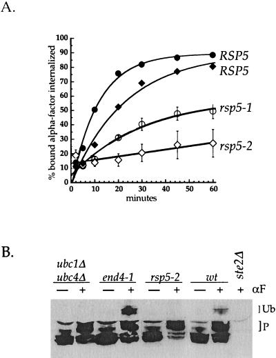
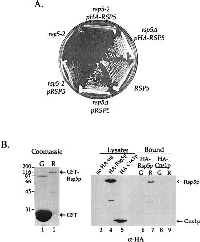
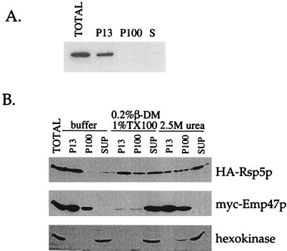
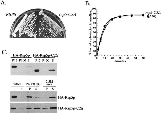
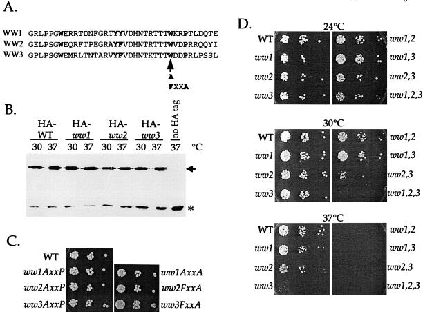
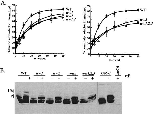
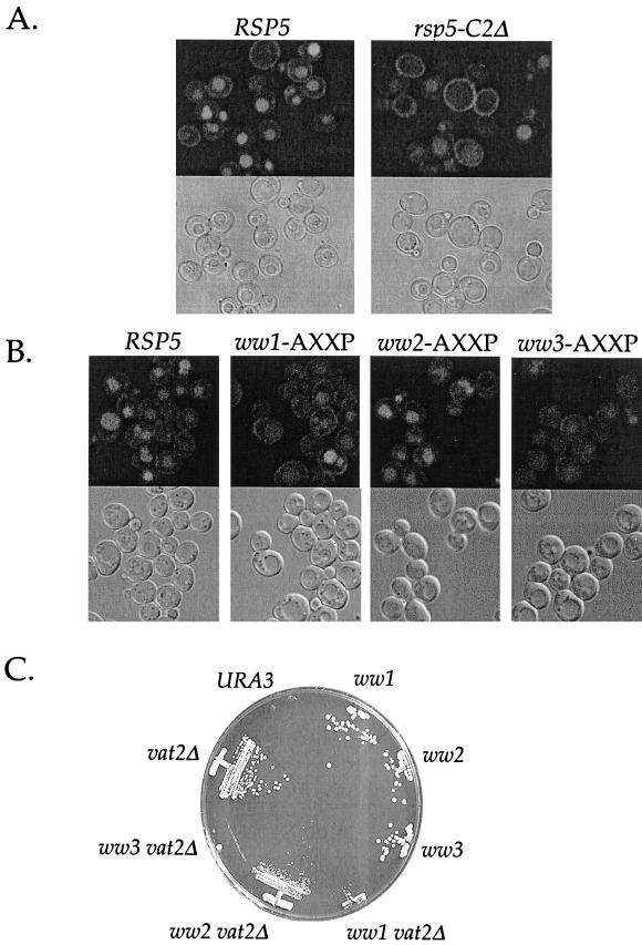
Yeast Rsp5p and its mammalian homologue, Nedd4, are hect domain ubiquitin-protein ligases (E3s) required for the ubiquitin-dependent endocytosis of plasma membrane proteins. Because ubiquitination is sufficient to induce internalization, E3-mediated ubiquitination is a key regulatory event in plasma membrane protein endocytosis. Rsp5p is an essential, multidomain protein containing an amino-terminal C2 domain, three WW protein-protein interaction domains, and a carboxy-terminal hect domain that carries E3 activity. In this study, we demonstrate that Rsp5p is peripherally associated with membranes and provide evidence that Rsp5p functions as part of a multimeric protein complex. We define the function of Rsp5p and its domains in the ubiquitin-dependent internalization of the yeast alpha-factor receptor, Ste2p. Temperature-sensitive rsp5 mutants were unable to ubiquitinate or to internalize Ste2p at the nonpermissive temperature. Deletion of the entire C2 domain had no effect on alpha-factor internalization; however, point mutations in any of the three WW domains impaired both receptor ubiquitination and internalization. These observations indicate that the WW domains play a role in the important regulatory event of selecting phosphorylated proteins as endocytic cargo. In addition, mutations in the C2 and WW1 domains had more severe defects on transport of fluid-phase markers to the vacuole than on receptor internalization, suggesting that Rsp5p functions at multiple steps in the endocytic pathway.


Figures

Figure 1
Figure 1
Rsp5p is required for Ste2p ubiquitination and internalization. (A) α-Factor internalization assays performed by the continuous presence protocol at 37°C (see MATERIALS AND METHODS). Cells were propagated in SD medium at 24°C and shifted to 37°C for 15 min before the addition of radiolabeled α-factor. RSP5 (LHY434, ♦), rsp5-2 (LHY433, ⋄), RSP5 (LHY291, ●), rsp5–1 (LHY23, ○). (B) Ste2p modification before and after α-factor (αF) treatment. ubc1Δ ubc4Δ (LHY1394), end4-1 (LHY501), rsp5-2 (LHY433), wild-type (LHY434), and ste2Δ (LHY10) cells were treated (+) or not (−) with 1 μM α-factor for 8 min at 37°C. Total cell lysates were resolved by SDS-PAGE, transferred to nitrocellulose, and probed with anti-Ste2p antibodies. Phosphorylated (P) and monoubiquitinated (Ub) receptor species are indicated by the labeled brackets.
Figure 2
Figure 2
Rsp5p self-interaction. (A) Semidominance of the rsp5-2 mutation. Cells of the indicated genotype were streaked onto YPUAD medium and grown for 2 d at 37°C. (B) GST-fusion protein precipitations. Left, Coomassie blue staining of the GST (G, lane 1) and GST-Rsp5p (R, lane 2) proteins used for each binding experiment. Bold arrows indicate the migration of GST and GST-Rsp5p. Molecular mass standards in kilodaltons are indicated at the left. Right, immunoblot analysis of GST pull-down experiment. Lysates prepared from LHY2066 (HA-RSP5) and MTY300 (HA-CNS1) cells were incubated with glutathione beads prebound to GST or to GST-Rsp5p. Bound proteins were eluted with Laemmli sample buffer. Eluted proteins and a fraction of the total cell lysates were resolved by SDS-PAGE, transferred to nitrocellulose, and probed with HA antibodies. Lanes 3–5, lysates of LHY2248 (no HA tag, lane 3), LHY2066 (HA-RSP5, lane 4), and MTY300 (HA-CNS1, lane 5) cells. Lanes 6–7, proteins from the LHY2066 lysate that bound to GST (lane 6) or GST-Rsp5p (lane 7). Lanes 8–9, proteins from the MTY300 lysate that bound to GST (lane 8) or GST-Rsp5p (lane 9). Arrows indicate the migration of HA-Rsp5p and HA-Cns1p.
Figure 3
Figure 3
Rsp5p associates with the membrane fraction of a cell lysate. (A) Differential centrifugation of a lysate prepared from cells expressing HA-Rsp5p (LHY2066). LHY2066 cells were propagated at 24°C in YPUA galactose medium; 13,000 × g (P13), 100,000 × g (P100), and soluble (S) cell fractions were resolved by SDS-PAGE, transferred to nitrocellulose, and probed with HA antibodies. (B) Differential centrifugation after biochemical treatments. LHY896 cells were grown in YPUAD medium at 30°C. The cell lysate was mock (buffer) treated, treated with 2.5 M urea, or treated with 1.0% (vol/vol) Triton X-100 (TX100) and 0.2% (wt/vol) dodecyl β-d-maltoside (β-DM) for 1 h on ice before fractionation. Fractions were analyzed as in A by immunoblotting with HA, c-myc, or hexokinase antibodies to detect HA-Rsp5p, myc-Emp47p, and hexokinase, respectively. An equivalent amount of the total cell extract is shown for comparison. SUP, supernatant.
Figure 4
Figure 4
Phenotypic analysis of rsp5-C2Δ cells. (A) Growth of RSP5 and rsp5-C2Δ cells. LHY1103 (RSP5) and LHY1101 (rsp5-C2Δ) cells were streaked onto YPUAD medium and grown for 2 d at 30°C. (B) α-Factor internalization assays performed by the pulse-chase protocol at 30°C (see MATERIALS AND METHODS). Cells were propagated in SD medium at 30°C. RSP5 (LHY1103, ●), rsp5-C2Δ (LHY1101, ○). (C) Fractionation of lysates prepared from cells expressing HA-Rsp5p or HA-Rsp5p-C2Δ. Top, differential centrifugation of LHY2066 (HA-Rsp5p) and LHY2232 (HA-Rsp5p-C2Δ) lysates. Cells were propagated in casamino acids-galactose medium at 24°C. Fractionation and immunoblot analysis were performed as described for Figure 3A. Bottom, fractionation of LHY1098 (HA-Rsp5p) and LHY1101 (HA-Rsp5p-C2Δ) cell lysates. Cells were propagated in YPUAD medium at 30°C. Lysates were fractionated into 100,000 × g pellet and supernatant fractions after biochemical treatment with 1.0% Triton X-100 (TX100), 2.5 M urea, or buffer as described for Figure 3.
Figure 5
Figure 5
Growth phenotypes of rsp5-ww domain mutants. (A) Sequence alignment of Rsp5p WW domains. WW1, amino acids 229–266. WW2, amino acids 331–368. WW3, amino acids 387–424. The highly conserved residues of the WW domain are in bold type. The arrow indicates the position of AXXP and FXXA mutations. (B) Expression of HA epitope-tagged ww-AXXP mutant proteins in cells expressing the mutant protein as the sole source of Rsp5p. Cells of the indicated genotypes were grown to early logarithmic phase in SD medium at 24°C. Cells were transferred to YPUAD medium, and aliquots of the cultures were shifted to 30 or 37°C for 15 min. Extracts prepared from these cells were prepared and analyzed by immunoblotting with HA antibodies. The asterisk indicates an unrelated cross-reacting band that serves as a loading control. WT, wild type. (C) ww-AXXP and ww-FXXA mutants are viable. Equal numbers of rsp5Δ cells carrying the indicated RSP5 plasmids were plated onto YPUAD medium (three serial 1:10 dilutions) and grown 3 d at 30°C. (D) Growth of single, double, and triple ww-AXXP mutants. Cells of the indicated genotype were serially diluted as in C and were grown on YPUAD medium at 24°C for 3 d, 30°C for 2 d, or 37°C for 2 d.
Figure 6
Figure 6
Rsp5p WW domains are required for receptor ubiquitination and internalization. (A) Continuous presence α-factor internalization assays. Cells were grown at 24°C to early logarithmic phase in SD medium and shifted to 37°C for 15 min before the addition of 35S-labeled α-factor. Wild-type (WT) (LHY1654, ●), rsp5-ww1 (LHY1729, ⋄), rsp5-ww2 (LHY1734, □), rsp5-ww1,2 (LHY2096, ○), rsp5-ww3 (LHY1735, ▵), rsp5-ww1,2,3 (LHY2098, ♦). (B) Ste2p modification before and after α-factor treatment. Wild-type (LHY2161), ww1 (LHY2162), ww2 (LHY2163), ww3 (LHY2164), ww1,2,3 (LHY2168), and rsp5-1 cells (LHY2169) expressing Ste2p from a galactose-inducible promoter were grown to early logarithmic phase in SD medium containing 2% galactose at 24°C. Cells were shifted to 37°C for 15 min and then treated (+) or not (−) with 1 μM α-factor for 8.5 min. Lysates were resolved by SDS-PAGE, transferred to nitrocellulose, and probed with anti-Ste2p antibodies. Labeled brackets indicate the migration of hyperphosphorylated (P) and ubiquitinated (Ub) species.
Figure 7
Figure 7
Fluid-phase endocytosis in rsp5 domain mutants. (A) LY uptake in RSP5 (LHY2066) and rsp5-C2Δ (LHY2232) cells. Cells were grown to early logarithmic phase at 30°C in YPUAD medium and then incubated with LY for 1 h. (B) LY uptake in ww-AXXP mutants. RSP5 (LHY1654), ww1 (LHY1729), ww2 (LHY1734), and ww3 (LHY1735) cells were grown to early logarithmic phase in YPUAD at 24°C, shifted to 30°C for 15 min, and then incubated for 75 min with LY at 30°C. Top panels are fluorescein isothiocyanate images. Bottom panels are differential interference contrast images of the same field of cells. (C) Synthetic lethality of ww1-AXXP and ww3-AXXP mutants with vat2Δ. ww-AXXP and ww-AXXP vat2Δ mutant cells carrying pRSP5(WT)[URA3] were grown on YNB medium containing 5-FOA. The growth of URA3 cells and cells carrying a disruption in VAT2 alone (vat2Δ) are shown for comparison.

References

    1. Babst M, Odorizzi G, Estepa EJ, Emr SD. Mammalian tumor susceptibility gene 101 (TSG101) and the yeast homologue, Vps23p, both function in late endosomal trafficking. Traffic. 2000;1:248–258. - PubMed
    1. Beck T, Schmidt A, Hall MN. Starvation induces vacuolar targeting and degradation of the tryptophan permease in yeast. J Cell Biol. 1999;146:1227–1238. - PMC - PubMed
    1. Bedford MT, Sarbassova D, Xu J, Leder P, Yaffe MB. A novel Pro-Arg motif recognized by WW domains. J Biol Chem. 2000;275:10359–10369. - PubMed
    1. Bénédetti H, Raths S, Crausaz F, Riezman H. The END3 gene encodes a protein that is required for the internalization step of endocytosis and for actin cytoskeleton organization in yeast. Mol Biol Cell. 1994;5:1023–1037. - PMC - PubMed
    1. Bonifacino JS, Weissman AM. Ubiquitin and the control of protein fate in the secretory and endocytic pathways. Annu Rev Cell Dev Biol. 1998;14:19–57. - PMC - PubMed

Publication types

MeSH terms