

Control of hair growth and follicle size by VEGF-mediated angiogenesis
- PMID: 11181640
- PMCID: PMC199257
- DOI: 10.1172/JCI11317
Control of hair growth and follicle size by VEGF-mediated angiogenesis
Abstract
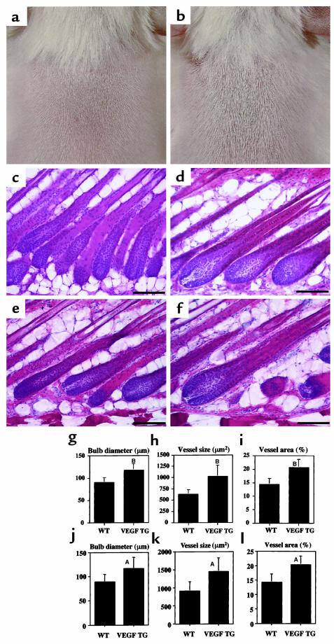
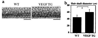
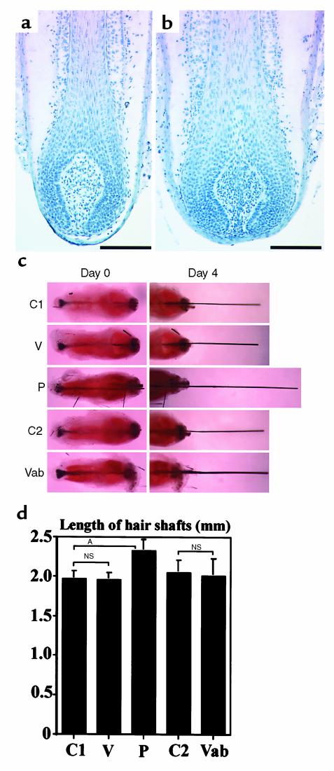
The murine hair follicle undergoes pronounced cyclic expansion and regression, leading to rapidly changing demands for its vascular support. Our study aimed to quantify the cyclic changes of perifollicular vascularization and to characterize the biological role of VEGF for hair growth, angiogenesis, and follicle cycling. We found a significant increase in perifollicular vascularization during the growth phase (anagen) of the hair cycle, followed by regression of angiogenic blood vessels during the involution (catagen) and the resting (telogen) phase. Perifollicular angiogenesis was temporally and spatially correlated with upregulation of VEGF mRNA expression by follicular keratinocytes of the outer root sheath, but not by dermal papilla cells. Transgenic overexpression of VEGF in outer root sheath keratinocytes of hair follicles strongly induced perifollicular vascularization, resulting in accelerated hair regrowth after depilation and in increased size of hair follicles and hair shafts. Conversely, systemic treatment with a neutralizing anti-VEGF antibody led to hair growth retardation and reduced hair follicle size. No effects of VEGF treatment or VEGF blockade were observed in mouse vibrissa organ cultures, which lack a functional vascular system. These results identify VEGF as a major mediator of hair follicle growth and cycling and provide the first direct evidence that improved follicle vascularization promotes hair growth and increases hair follicle and hair size.
Figures


Similar articles
-
Expression of vascular endothelial growth factor (VEGF) in various compartments of the human hair follicle.Arch Dermatol Res. 1998 Dec;290(12):661-8. doi: 10.1007/s004030050370. Arch Dermatol Res. 1998. PMID: 9879835
-
Thrombospondin-1 plays a critical role in the induction of hair follicle involution and vascular regression during the catagen phase.J Invest Dermatol. 2003 Jan;120(1):14-9. doi: 10.1046/j.1523-1747.2003.12045.x. J Invest Dermatol. 2003. PMID: 12535193
-
[Pilar growth: VEGF and fibroblasts of the follicular papilla].Ann Dermatol Venereol. 1998 Nov;125 Suppl 2:S9-11. Ann Dermatol Venereol. 1998. PMID: 9922881 French. No abstract available.
-
[Implication of VEGF, steroid hormones and neuropeptides in hair follicle cell responses].Ann Dermatol Venereol. 2002 May;129(5 Pt 2):783-6. Ann Dermatol Venereol. 2002. PMID: 12223959 Review. French.
-
[Regulation of hair follicle cycle].Morfologiia. 2014;146(5):83-7. Morfologiia. 2014. PMID: 25823297 Review. Russian.
Cited by
-
Hair Regeneration Treatment Using Adipose-Derived Stem Cell Conditioned Medium: Follow-up With Trichograms.Eplasty. 2015 Mar 26;15:e10. eCollection 2015. Eplasty. 2015. PMID: 25834689 Free PMC article.
-
Therapeutic potential of gingival fibroblasts for cutaneous radiation syndrome: comparison to bone marrow-mesenchymal stem cell grafts.Stem Cells Dev. 2015 May 15;24(10):1182-93. doi: 10.1089/scd.2014.0486. Epub 2015 Feb 26. Stem Cells Dev. 2015. PMID: 25584741 Free PMC article.
-
Effectiveness of the combinational treatment of Laminaria japonica and Cistanche tubulosa extracts in hair growth.Lab Anim Res. 2015 Mar;31(1):24-32. doi: 10.5625/lar.2015.31.1.24. Epub 2015 Mar 20. Lab Anim Res. 2015. PMID: 25806080 Free PMC article.
-
An important role of podoplanin in hair follicle growth.PLoS One. 2019 Jul 23;14(7):e0219938. doi: 10.1371/journal.pone.0219938. eCollection 2019. PLoS One. 2019. PMID: 31335913 Free PMC article.
-
Sostdc1 Secreted from Cutaneous Lymphatic Vessels Acts as a Paracrine Factor for Hair Follicle Growth.Curr Issues Mol Biol. 2022 May 12;44(5):2167-2174. doi: 10.3390/cimb44050146. Curr Issues Mol Biol. 2022. PMID: 35678675 Free PMC article.
References
-
- Chase HH. Growth of the hair. Physiol Rev. 1954; 34:113–126. - PubMed
-
- Hardy MH. The secret life of the hair follicle. Trends Genet. 1992; 8:55–60. - PubMed
-
- Paus R, Cotsarelis G. The biology of hair follicles. N Engl J Med. 1999; 341:491–497. - PubMed
-
- Montagna W, Ellis RA. Histology and cytochemistry of human skin. XIII. The blood supply of the hair follicle. J Natl Cancer Inst. 1957; 19:451–463. - PubMed
-
- Durward, A., and Rudall, K.M. 1958. The vascularity and patterns of growth of hair follicles. In The biology of hair growth. W. Montagna and R.A. Ellis, editors. Academic Press. New York, New York, USA. 189–218.
Publication types
MeSH terms
Substances
Grants and funding
LinkOut - more resources
Full Text Sources
Other Literature Sources
Molecular Biology Databases
