. 2001 Feb;107(4):419-30.
doi: 10.1172/JCI8797.

Increased atherosclerosis in myeloperoxidase-deficient mice

Affiliations

Increased atherosclerosis in myeloperoxidase-deficient mice

M L Brennan et al. J Clin Invest. 2001 Feb.

Abstract

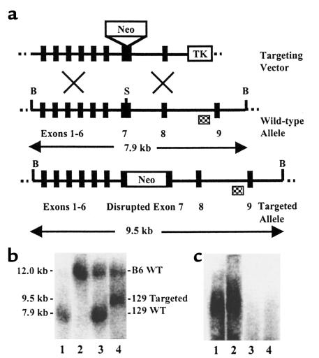
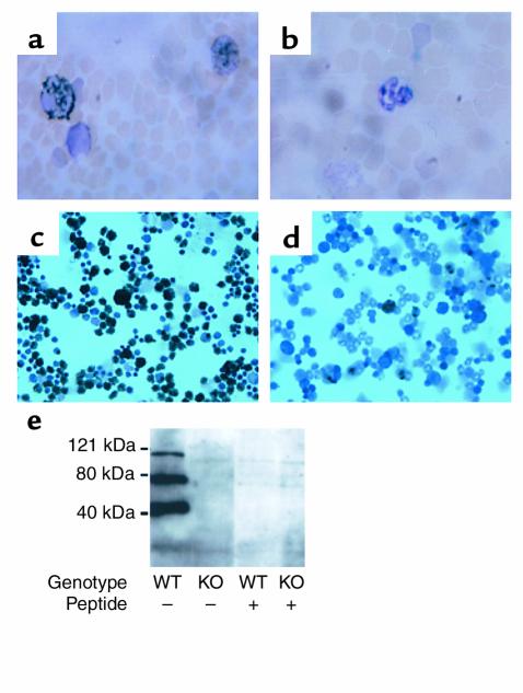
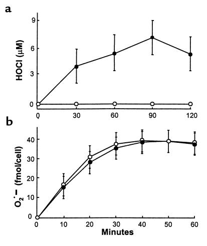
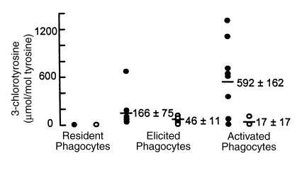
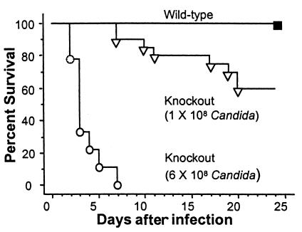
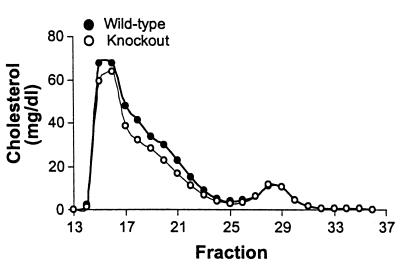
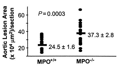
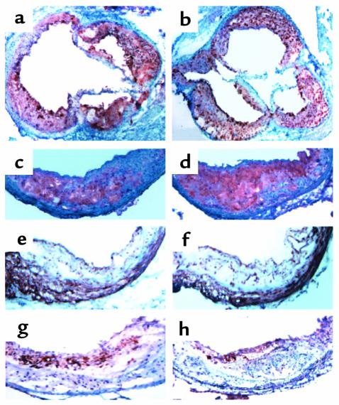
Myeloperoxidase (MPO), a heme enzyme secreted by activated phagocytes, generates an array of oxidants proposed to play critical roles in host defense and local tissue damage. Both MPO and its reaction products are present in human atherosclerotic plaque, and it has been proposed that MPO oxidatively modifies targets in the artery wall. We have now generated MPO-deficient mice, and show here that neutrophils from homozygous mutants lack peroxidase and chlorination activity in vitro and fail to generate chlorotyrosine or to kill Candida albicans in vivo. To examine the potential role of MPO in atherosclerosis, we subjected LDL receptor-deficient mice to lethal irradiation, repopulated their marrow with MPO-deficient or wild-type cells, and provided them a high-fat, high-cholesterol diet for 14 weeks. White cell counts and plasma lipoprotein profiles were similar between the two groups at sacrifice. Cross-sectional analysis of the aorta indicated that lesions in MPO-deficient mice were about 50% larger than controls. Similar results were obtained in a genetic cross with LDL receptor-deficient mice. In contrast to advanced human atherosclerotic lesions, the chlorotyrosine content of aortic lesions from wild-type as well as MPO-deficient mice was essentially undetectable. These data suggest an unexpected, protective role for MPO-generated reactive intermediates in murine atherosclerosis. They also identify an important distinction between murine and human atherosclerosis with regard to the potential involvement of MPO in protein oxidation.


Figures

Figure 1
Figure 1
Targeted disruption of the mouse MPO gene. (a) The targeting vector, wild-type MPO locus (including exons 1–9), and targeted MPO gene are shown. Exon 7 was disrupted by insertion of positive-selection marker PGK-Neo (Neo) at a BssHII (S) site. HSV-thymidine kinase (TK) served for negative selection. BglII (B) digestion sites, lengths of diagnostic BglII fragments, and external probe (checkered box) used for Southern blot analysis are indicated. (b) DNA isolated from progeny of chimeric founder male mated with C57BL/6J female was subjected to BglII digestion and Southern blot analysis. Lanes 1 and 2 contained the wild-type controls 129/SvJ DNA (lane 1) and C57BL/6J DNA (lane 2). Note the difference in band size between the two strains, indicative of a restriction fragment length variant. Strain 129/SvJ has BglII restriction enzyme sites at nucleotides 189 and 8075, yielding a 7.9-kb product, whereas C57BL/6J is lacking one of these sites, and the fragment generated represents digestion at a more distal BglII site. Restriction variants, such as this one, are due to naturally occurring nucleic acid sequence differences between inbred strains. Lanes 3 and 4 are from progeny. The larger band in both lanes is inherited from the C57BL/6J mother, and the lower band, inherited from the chimeric father, is either wild-type (lane 3) or targeted (lane 4). (c) RNA samples isolated from bone marrow from wild-type (lanes 1 and 2) and mutant (lanes 3 and 4) mice were subjected to Northern blot analysis with a human cDNA probe.
Figure 2
Figure 2
Functional ablation of the MPO gene. Peroxidase stains were performed on MPO+/+ (a) and MPO–/– (b) peripheral blood samples and MPO+/+ (c) and MPO–/– (d) bone marrow samples. Cell types were determined based on nuclear staining and size. Shown are peroxidase-positive eosinophil(s) (a, c, d), neutrophil(s) (a, c), and monocytes (c), and peroxidase-negative neutrophil(s) (b, d) and monocytes (d). Bone marrow cells from MPO+/+ and MPO–/– mice were subjected to SDS-PAGE. Western blot analysis (e) was performed with a rabbit polyclonal Ab against the carboxy-terminal 14 amino acids of MPO. The peptide immunogen was used in indicated lanes to block immunoreactivity. MPO is synthesized as a preproenzyme and undergoes processing. The three major bands in the wild-type mice likely represent prepro-MPO, processed (but unclipped) heavy and light chains of MPO, and the heavy chain of MPO. Microsatellites of molecular-weight-size markers are indicated.
Figure 3
Figure 3
Superoxide and hypochlorous acid production by peritoneal phagocytes in vitro. (a) Peritoneal exudate cells were isolated from thioglycollate-treated wild-type (filled circle) or MPO-deficient (open circle) animals (n = 4/genotype) and stimulated with phorbol ester, and HOCl production was measured as taurine chloramine formation. (b) Peritoneal exudate cells were isolated from thioglycollate-treated wild-type (filled circle) or MPO-deficient (open circle) LDLR-deficient animals (n = 6/genotype) and stimulated with phorbol ester, and superoxide (O2•–) production was quantitated as the reduction of cytochrome c.
Figure 4
Figure 4
Isotope-dilution GC/MS analysis of 3-chlorotyrosine in phagocytes isolated from MPO+/+ and MPO–/– mice. Resident peritoneal cells (resident phagocytes; n = 3), thioglycollate-elicited peritoneal exudate cells (quiescent phagocytes; MPO+/+, n = 8, and MPO–/–, n = 10), and thioglycollate-elicited, zymosan-stimulated peritoneal exudate cells (activated phagocytes; MPO+/+, n = 8, and MPO–/–, n = 6) were isolated by lavage from wild-type (filled circle) and MPO-deficient (open circle) animals. After acid hydrolysis and derivatization, 3-chlorotyrosine levels of cellular proteins were determined by negative-ion electron capture GC/MS with selected ion monitoring. Results are normalized to tyrosine, the precursor amino acid.
Figure 5
Figure 5
Survival of MPO+/+ and MPO–/– mice after intraperitoneal challenge with Candida. Each mouse was injected with 108 CFU of C. albicans (MPO+/+, n = 19; MPO–/–, n = 19) or 6 × 10⁸ CFU of C. albicans (MPO+/+, n = 9; MPO–/–, n = 9). Survival was monitored for 65 days, and no mortality occurred after 20 days. Results are from two independent experiments.
Figure 6
Figure 6
FPLC fractionation and cholesterol determination of plasma from MPO wild-type/LDLR-deficient and MPO-deficient/LDLR-deficient animals. Mice were fed an atherogenic diet for 14 weeks. After a 16-hour fast, they were bled, equal amounts of plasma from eight animals in each group were pooled, and FPLC was performed. Total cholesterol was determined in fractions as described (65).
Figure 7
Figure 7
Effect of MPO deficiency on atherosclerosis development in mice. LDLR-deficient mice that were also MPO wild-type or deficient were fed an atherogenic diet for 14 weeks, and atherosclerosis was assessed. The total lesion areas in sections from the proximal aorta of LDLR-deficient mice containing the wild-type or targeted MPO allele were determined using oil red O staining. Values for individual animals (n = 21 per group) are shown.
Figure 8
Figure 8
Atherosclerosis in MPO wild-type/LDLR-deficient and MPO-deficient/LDLR-deficient animals. Cross-sections through the aortic root were stained with oil red O to detect neutral lipid in wild-type (a) and MPO-deficient (b) animals. Extensive lesions are visible. Immunohistochemical stains for monocyte/macrophages in wild-type (c) and MPO-deficient (d) animals are shown with intense red staining. Smooth muscle cell staining in wild-type (e) and MPO-deficient (f) lesions is detected with dark brown staining and was similar between groups. Staining with Ab’s against MPO in wild-type (g) or MPO-deficient (h) animals showed cross-reactivity.

Comment in

  • The proper study of mankind.
    Nauseef WM. Nauseef WM. J Clin Invest. 2001 Feb;107(4):401-3. doi: 10.1172/JCI11713. J Clin Invest. 2001. PMID: 11181638 Free PMC article. No abstract available.

References

    1. Agner K. Verdoperoxidase: a ferment isolated from leukocytes. Acta Physiol Scand. 1941; 2:1–62.
    1. Babior BM. Oxygen-dependent microbial killing by phagocytes. N Engl J Med. 1978; 298:659–666. - PubMed
    1. Klebanoff, S.J., and Clark, R.A. 1978. The neutrophil: function and clinical disorders. Elsevier/North Holland Biomedical Press. Amsterdam, The Netherlands. 1–810.
    1. Weiss SJ. Tissue destruction by neutrophils. N Engl J Med. 1989; 320:365–376. - PubMed
    1. Shultz J, Kaminker K. Myeloperoxidase of the leucocyte of normal human blood. 1. Content and localization. Arch Biochem Biophys. 1962; 96:465–467. - PubMed

Publication types