. 2001 Feb;107(4):519-28.
doi: 10.1172/JCI8027.

Rheumatoid arthritis synovial T cells regulate transcription of several genes associated with antigen-induced anergy

Affiliations

Rheumatoid arthritis synovial T cells regulate transcription of several genes associated with antigen-induced anergy

M Ali et al. J Clin Invest. 2001 Feb.

Abstract

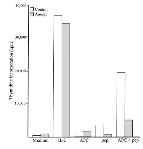
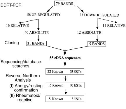
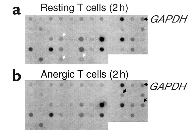
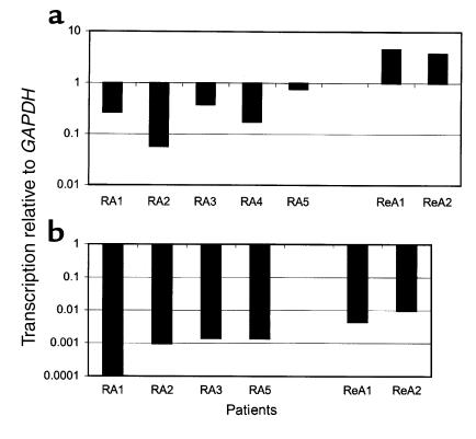
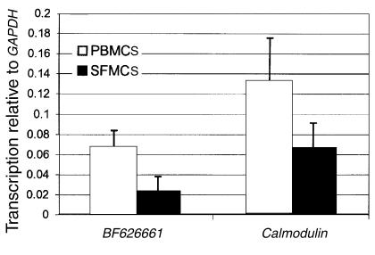
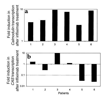
Rheumatoid arthritis (RA) is a chronic, inflammatory synovitis whose pathogenesis may involve autoimmune mechanisms. Anergy is a state of T-cell nonresponsiveness characterized by downregulated IL-2 production. Paradoxically, RA T cells are hyporesponsive and proliferate poorly to antigens and mitogens, thus sharing some characteristics with anergic T cells. We analyzed the molecular basis of anergy in cloned human CD4+ T cells using differential display RT-PCR and subsequently examined the levels of differentially expressed transcripts in RA and, as control, reactive arthritis (ReA) synovium. Several transcriptional events were common to anergic T cells and RA synovium. These included downregulation of CALMODULIN:, which is critical to T-cell activation, and of cellular apoptosis susceptibility protein, which may mediate resistance to apoptosis in RA. Transcription of CALMODULIN: in RA synovium was less than 1% of that in ReA and was lower in RA synovial fluid mononuclear cells than in paired PBMCs. Following anti-TNF-alpha therapy in vivo, RA PBMC CALMODULIN: transcripts increased five- to tenfold. Pharmacological calmodulin blockade in vitro impaired antigen-specific proliferation. These data provide a link between reduced CALMODULIN: transcription and impaired T-cell responsiveness in RA. The identification of transcriptional changes common to anergic and RA synovial T cells should help interpret some of the characteristic RA cellular defects.


Figures

Figure 1
Figure 1
Induction of anergy in the human CD4+ T-cell clone HA1.7. HA1.7 (106/ml) was incubated with peptide HA 306-318 (50 μg/ml) for 24 hours or with medium alone. The anergic state of the cells was subsequently tested by an immunogenic challenge. Peptide-preincubated cells (dark bars, 104/well) did not proliferate when stimulated with HA 306-318 in the presence of APC, but their viability was confirmed by responsiveness to exogenous IL-2. In contrast, control cells (light bars, 104/well) proliferated after either antigenic challenge or IL-2. Neither control nor anergic cells proliferated when incubated with medium, APC, or peptide alone. The figure shows proliferation after 48 hours of stimulation, measured as [3H]thymidine incorporation.
Figure 2
Figure 2
A schematic of the DDRT-PCR and confirmatory analysis. After differential display (DDRT-PCR) analysis of T cells undergoing anergy induction, 55 distinct cDNA sequences were isolated. These formed the basis of the array that was used to confirm differential gene expression and compare transcription in RA and ReA synovium.
Figure 3
Figure 3
Confirmation of DDRT-PCR results by reverse Northern blotting analysis. Replicate nylon-filter membranes were prepared, comprising 200-bp cDNA fragments (2 μg of each) representing each differentially expressed transcript. Radiolabeled cDNA probes were prepared from 5 μg total RNA extracted from (a) resting or (b) anergic T cells. The probes were hybridized to the filters overnight, and, after extensive washing, the filters were exposed for 3 days to Phosphor Screens. Transcripts downregulated during anergy induction are highlighted by white arrows. Transcripts upregulated are highlighted by black arrows. The blot represents all 55 transcripts that were considered to be differentially expressed after the initial DDRT-PCR screen, as well as the housekeeping control transcript, GAPDH.
Figure 4
Figure 4
Calmodulin and CAS transcription in individual biopsies. First-strand cDNA was prepared from synovial tissue from individuals with RA and with ReA. Real-time PCR was used to quantitate transcription of (a) Calmodulin and (b) CAS relative to the housekeeping transcript GAPDH. P = 0.002 for Calmodulin and 0.035 for CAS, for RA vs. ReA (unpaired t test).
Figure 5
Figure 5
Quantitation of gene transcription in mononuclear cells. First-strand cDNA was prepared from PBMCs and SFMCs from RA patients. Real-time PCR was used to quantitate transcription of Calmodulin and BF626661 relative to GAPDH as a housekeeping transcript. The figure shows the ratio of target gene to GAPDH transcription in SFMCs and PBMCs (mean and SD of four patients). P = 0.050 for Calmodulin and 0.026 for BF626661 for PBMC vs. SFMC (paired t test).
Figure 6
Figure 6
Transcription of Calmodulin and CAS in PBMCs isolated from RA patients before and after treatment with anti–TNF-α mAb. First-strand cDNA was prepared from PBMCs from six patients before and 2 weeks after receiving 3 mg/kg Infliximab. Real-time PCR was used to quantitate transcription of (a) Calmodulin and (b) CAS relative to GAPDH. The figure shows the ratio of transcripts in the posttreatment samples relative to the pretreatment samples for each patient.
Figure 7
Figure 7
Inhibition of antigen-specific PBMC proliferation by calmodulin blockade. PBMCs were prepared from normal controls. Proliferation assays were established using PPD as a test antigen in the presence or absence of TFP, a pharmacological antagonist of calmodulin. The figure illustrates pooled data for four individuals of the percentage inhibition (mean ± SD) of the proliferation index at each dose of TFP. The proliferation index was calculated as the ratio of counts between stimulated and unstimulated cells.

References

    1. Van Boxel JA, Paget SA. Predominantly T cell infiltrate in rheumatoid synovial membranes. N Engl J Med. 1975; 293:517–520. - PubMed
    1. Fox DA. The role of T-cells in the immunopathogenesis of rheumatoid arthritis. Arthritis Rheum. 1997; 40:598–609. - PubMed
    1. Cush JJ, Lipsky PE. Phenotypic analysis of synovial and peripheral blood lymphocytes isolated from patients with rheumatoid arthritis. Arthritis Rheum. 1988; 31:1230–1238. - PubMed
    1. Silverman HA, Johnson JS, Vaughan JH, McGlamory JC. Altered lymphocyte reactivity in rheumatoid arthritis. Arthritis Rheum. 1976; 19:509–515. - PubMed
    1. Salmon M, et al. Inhibition of T cell apoptosis in the rheumatoid synovium. J Clin Invest. 1997; 99:439–446. - PMC - PubMed

Publication types