. 2001 Feb 19;193(4):417-26.
doi: 10.1084/jem.193.4.417.

IkappaB kinase alpha is essential for mature B cell development and function

Affiliations

IkappaB kinase alpha is essential for mature B cell development and function

T Kaisho et al. J Exp Med. .

Abstract

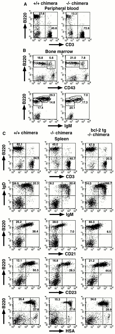
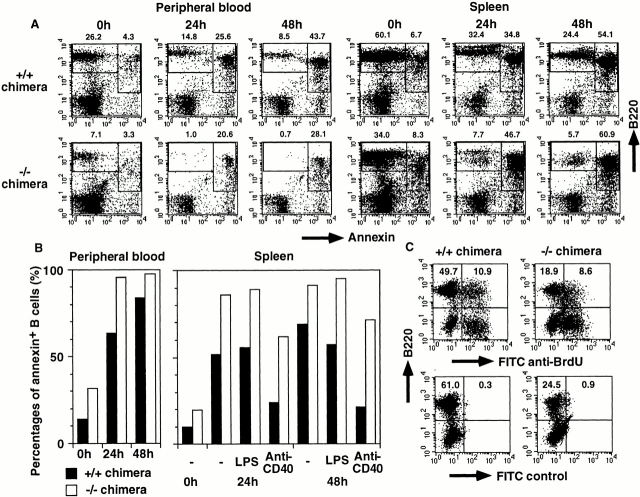
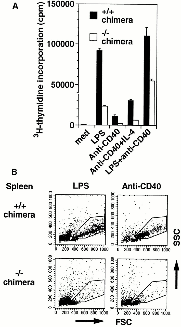
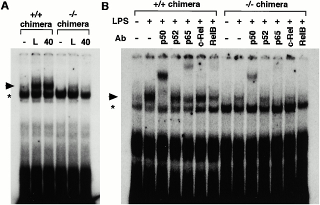
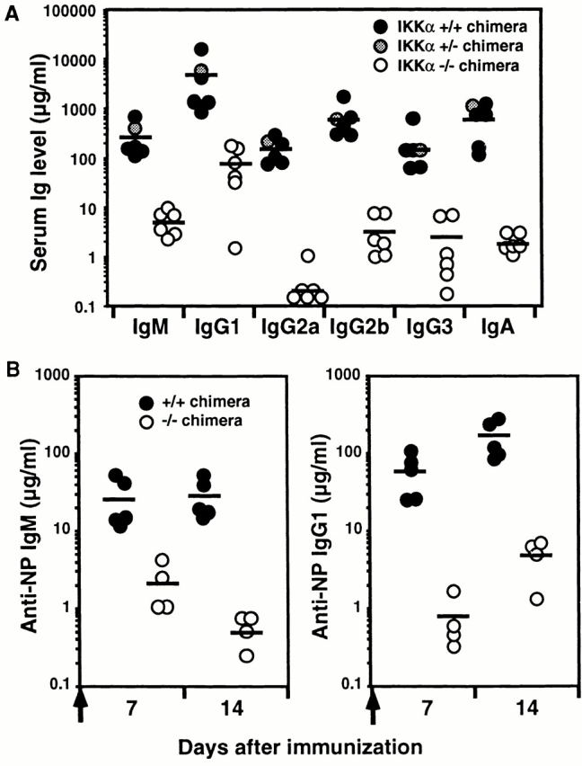
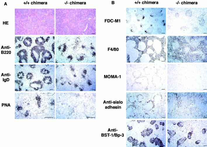
IkappaB kinase (IKK) alpha and beta phosphorylate IkappaB proteins and activate the transcription factor, nuclear factor (NF)-kappaB. Although both are highly homologous kinases, gene targeting experiments revealed their differential roles in vivo. IKKalpha is involved in skin and limb morphogenesis, whereas IKKbeta is essential for cytokine signaling. To elucidate in vivo roles of IKKalpha in hematopoietic cells, we have generated bone marrow chimeras by transferring control and IKKalpha-deficient fetal liver cells. The mature B cell population was decreased in IKKalpha(-/-) chimeras. IKKalpha(-/-) chimeras also exhibited a decrease of serum immunoglobulin basal level and impaired antigen-specific immune responses. Histologically, they also manifested marked disruption of germinal center formation and splenic microarchitectures that depend on mature B cells. IKKalpha(-/-) B cells not only showed impairment of survival and mitogenic responses in vitro, accompanied by decreased, although inducible, NF-kappaB activity, but also increased turnover rate in vivo. In addition, transgene expression of bcl-2 could only partially rescue impaired B cell development in IKKalpha(-/-) chimeras. Taken together, these results demonstrate that IKKalpha is critically involved in the prevention of cell death and functional development of mature B cells.


Figures

Figure 1
Figure 1
Mature B cell decrease in IKKα2/− RAG2-deficient B6 chimeras. Single cell suspensions from (A) PB, (B) BM, and (C) spleen were stained with the indicated Abs and analyzed using a FACSCalibur™ with CELLQuest™ software. The percentages of the quadrants or enclosed areas are indicated by numbers. For BM, triple color analysis was performed, and CD43 versus B220 and IgM versus B220 profiles are shown for IgM and CD43 lymphoid cells, respectively. In C, data from IKKα2/− chimeras with transgene expression of bcl-2 are also shown. Four independent experiments were performed with similar results. One representative experiment is shown.
Figure 2
Figure 2
Impaired survival in vitro and increased turnover in vivo of IKKα2/− B cells. (A and B) PB and splenocytes from IKKα1/+ and IKKα2/− RAG2-deficient B6 chimeras were cultured in complete RPMI 1640 in the absence or presence of mitogens for the indicated periods and stained with FITC–annexin V and PE-B220. Percentages of annexin+ B cells were calculated by dividing percentages of B220+annexin+ cells by those of total B (B220+annexin+ and B220+annexin) cells and shown as bar graphs in B. (C) Increased B cell turnover in IKKα2/− chimeras. The turnover of splenic B cells was determined by BrdU incorporation. Numbers represent percentages of the quadrants. Experiments were independently performed three times with similar results. One representative experiment is shown.
Figure 3
Figure 3
Impaired mitogenic responses of IKKα2/− B cells. (A) Splenic B cells from IKKα1/+ and IKKα2/− RAG2-deficient B6 chimeras were cultured in the absence or presence of mitogens for 72 h and analyzed for their [3H]thymidine incorporation. The data indicate means ± SD of triplicate samples of one representative experiment. (B) Splenocytes from IKKα1/+ and IKKα2/− RAG2-deficient B6 chimeras were cultured with LPS or anti-CD40 for 48 h. Forward scatter (FSC) and side scatter (SSC) of harvested cells were analyzed by FACS®. Blasts are indicated by enclosed areas. Experiments were independently performed three times with similar results. One representative experiment is shown.
Figure 4
Figure 4
NF-κB DNA binding activity in IKKα1/+ and IKKα2/− B cells. (A) Splenic B cells were stimulated with or without 25 μg/ml LPS (L) or 0.5 μg/ml anti-CD40 40 for 1.5 h. The whole cell lysates were prepared and NF-κB activity was determined by electrophoretic mobility shift assay. (B) LPS-induced NF-κB complexes were analyzed by supershift analysis. Specific NF-κB complexes are indicated by arrowheads. *Nonspecific binding. Similar results were obtained from three independent experiments.
Figure 5
Figure 5
Decrease of basal A1 gene expression in IKKα2/− B cells. A1 expression was analyzed in IKKα1/+ or IKKα2/− B cells with or without LPS (L) or anti-CD40 40 stimulation through reverse transcription PCR analysis. Basal A1 gene expression was determined by amplification with 30 cycles. As controls, data for β-actin expression are shown. M, Φ×174/HaeIII digest marker.
Figure 6
Figure 6
Decreased Ig production and impaired T cell–dependent immune responses in IKKα2/− chimeras. (A) Serum Ig isotype levels in unimmunized, IKKα1/+, IKKα1/−, and IKKα2/− RAG2-deficient B6 chimeras. Serum Ig titers were determined by isotype-specific ELISA. (B) Immune responses to the T cell–dependent Ag, NP-CG. IKKα1/+ and IKKα2/− RAG2-deficient B6 chimeras were immunized with alum-precipitated NP-CG and bled at the indicated days. NP-specific IgM and IgG1 titers were determined by ELISA. Bars indicate mean values.
Figure 7
Figure 7
(A) Impaired GC formation in IKKα2/− chimeras. IKKα1/+ and IKKα2/− RAG2-deficient B6 chimeras were immunized with alum-precipitated NP-CG and killed 14 d after injection. Splenic sections were stained with HE or immunostained with B220, anti-IgD, or PNA. (B) Impairment of splenic microarchitecture in IKKα2/− chimeras. Frozen sections were stained with FDC-M1, F4/80, MOMA-1, or antisialoadhesin mAbs or rabbit anti–BST-1/Bp-3 antiserum. Scale bars, 200 μm.

References

    1. Baeuerle P.A., Baltimore D. NF-κBten years after. Cell. 1996;87:13–20. - PubMed
    1. Barnes P.J., Karin M. Nuclear factor-κBa pivotal transcription factor in chronic inflammatory diseases. N. Engl. J. Med. 1997;336:1066–1071. - PubMed
    1. Regnier C.H., Song H.Y., Gao X., Goeddel D.V., Cao Z., Rothe M. Identification and characterization of an IκB kinase. Cell. 1997;90:373–383. - PubMed
    1. Zandi E., Rothwarf D.M., Delhase M., Hayakawa M., Karin M. The IκB kinase complex (IKK) contains two kinase subunits, IKKα and IKKβ, necessary for IκB phosphorylation and NF-κB activation. Cell. 1997;91:243–252. - PubMed
    1. Woronicz J.D., Gao X., Cao Z., Rothe M., Goeddel D.V. IκB kinase-βNF-κB activation and complex formation with IκB kinase-α and NIK. Science. 1997;278:866–869. - PubMed

Publication types